Collateral ligament (CL) injury - aftercare
Medial collateral ligament injury - aftercare; MCL injury - aftercare; Lateral collateral ligament injury - aftercare; LCL injury - aftercare
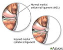
A ligament is a band of tissue that connects bone to bone. The collateral ligaments are located on the outside of your knee joint. They help connect the bones of your upper and lower leg, inside your knee joint.
- The lateral collateral ligament (LCL) runs on the outer side of your knee.
- The medial collateral ligament (MCL) runs along the inside of your knee.
A collateral ligament injury occurs when the ligaments are stretched or torn. A partial tear occurs when only part of the ligament is torn. A complete tear occurs when the entire ligament is torn into two pieces.
More About Your Injury
The collateral ligaments help keep your knee stable. They help keep your leg bones in place and keep your knee from moving too far sideways.
A collateral ligament injury can occur if you get hit very hard on the inside or outside of your knee.
Skiers and people who play basketball, football, or soccer are more likely to have this type of injury.
What to Expect
With a collateral ligament injury, you may notice:
- Your knee is unstable and can shift side to side as if it "gives way"
- Locking or catching of the knee with movement
- Knee swelling
-
Knee pain
along the inside or outside of your knee
Knee pain
Knee pain is a common symptom in people of all ages. It may start suddenly, often after an injury or exercise. Knee pain also may begin as a mild d...
 ImageRead Article Now Book Mark Article
After examining your knee, your doctor may send you to have an MRI . An MRI is a device that can take pictures of the tissues around your knee. The pictures will show whether these tissues have been stretched or torn. You also may have an x-ray to see if there is any damage to the bones in your knee.
MRI
A knee MRI (magnetic resonance imaging) scan uses energy from strong magnets to create pictures of the knee joint and muscles and tissues. An MRI doe...
x-ray
This test is an x-ray of a knee, shoulder, hip, wrist, ankle, or other joint.
If you have a collateral ligament injury, you may need:
- Crutches to walk until the swelling and pain get better
- A brace to support and stabilize your knee
- Physical therapy to help improve joint motion and leg strength
Most people do not need surgery for an MCL injury. However, you may need surgery if your LCL is injured or if your injuries are severe and involve other ligaments in your knee.
Self-care at Home
Follow R.I.C.E. to help reduce pain and swelling:
- Rest your leg. Avoid putting weight on it.
- Ice your knee for 20 minutes at a time, 3 to 4 times a day.
- Compress the area by wrapping it with an elastic bandage or compression wrap.
- Elevate your leg by raising it above the level of your heart.
You can use ibuprofen (Advil, Motrin), or naproxen (Aleve, Naprosyn) to reduce pain and swelling. Acetaminophen (Tylenol) helps with pain, but not swelling. You can buy these pain medicines at the store.
- Talk with your doctor before using these medicines if you have heart disease, high blood pressure, kidney disease, or have had stomach ulcers or internal bleeding in the past.
- DO NOT take more than the amount recommended on the bottle or by your doctor.
Activity
You should not put all of your weight on your leg if it hurts, or if your doctor tells you not to. Rest and self-care may be enough to allow the tear to heal. You should use crutches to protect the injured ligament.
Afterward, you will learn exercises to make the muscles, ligaments, and tendons around your knee stronger and more flexible.
- You may need to work with a physical therapist to regain knee and leg strength.
- Slowly, you can return to normal activities and perhaps return to sports again. Ask your doctor.
When to Call the Doctor
Call your doctor if:
- You have increased swelling or pain
- Self-care does not seem to help
- You lose feeling in your foot
- Your foot and leg feels cold or changes color
If you have surgery, call the doctor if you have:
- A fever of 100°F (38°C) or higher
- Any drainage from the injection or incision site
- Bleeding that will not stop
References
Lento P, Akuthota V. Collateral ligament sprain. In: Frontera WR, Silver JK, Rizzo TD Jr, eds. Essentials of Physical Medicine and Rehabilitation: Musculoskeletal Disorders, Pain, and Rehabilitation . 3rd ed. Philadelphia, PA: Elsevier Saunders; 2015:chap 66.
Miller III RH, Azar, FM. Knee injuires. In: Canale ST, Beaty JH, Daugherty K, Jones L, et al. Canale & Beaty: Campbell's Operative Orthopaedics . 12th ed. Philadelphia, PA: Elsevier Mosby; 2013:chap 45.
Niska JA, Petrigliano FA, McAllister Dr. Anterior cruciate ligament injuries (including revision). In: Miller MD, Thompson SR, eds. DeLee and Drez's Orthopaedic Sports Medicine: Principles and Practice . 4th ed. Philadelphia, PA: Elsevier Saunders; 2015:chap 98.
Wilson BF, Johnson DL. Medial collateral ligament and posterior medial corner injuries. In: Miller MD, Thompson SR, eds. DeLee and Drez's Orthopaedic Sports Medicine: Principles and Practice . 4th ed. Philadelphia, PA: Elsevier Saunders; 2015:chap 100.
-
Medial collateral ligament - illustration
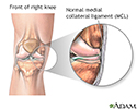
The medial collateral ligament connects the end of the femur (thigh) to the top of the tibia (shin bone). The medial collateral ligament provides stability against valgus stress. A valgus stress is described as a pressure applied to the leg that tries to bend the lower leg sideways at the knee, away from the other leg. Tackling in football or soccer are two common causes of this injury.
Medial collateral ligament
illustration
-
Knee pain - illustration
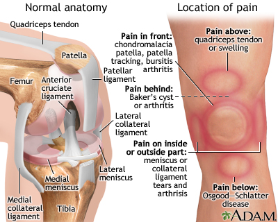
The location of knee pain can help identify the problem. Pain on the front of the knee can be due to bursitis, arthritis, or softening of the patella cartilage as in chondromalacia patella. Pain on the sides of the knee is commonly related to injuries to the collateral ligaments, arthritis, or tears to the meniscuses. Pain in the back of the knee can be caused by arthritis or cysts, known as Baker’s cysts. Baker’s cysts are an accumulation of joint fluid (synovial fluid) that forms behind the knee. Overall knee pain can be due to bursitis, arthritis, tears in the ligaments, osteoarthritis of the joint, or infection. Instability, or giving way, is also another common knee problem. Instability is usually associated with damage or problems with the meniscuses, collateral ligaments, or patella tracking.
Knee pain
illustration
-
Medial collateral ligament pain - illustration
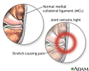
Initial treatment of an MCL injury includes ice to the area, elevation of the joint above the level of the heart, non-steroidal anti-inflammatory drugs (NSAIDs), and limited physical activity until the pain and swelling subside. A hinged knee immobilizer should be used to protect the ligament as it heals. The extent of this type of injury is usually excessive stretching of the ligament causing the pain and tenderness.
Medial collateral ligament pain
illustration
-
Medial collateral ligament injury - illustration
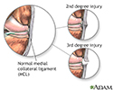
A second degree injury is a partial tear with no firm endpoint when the joint is stressed, and a third degree is a complete tear of the ligament. A physical examination will be done to test the extent of damage. Some other tests may include an MRI or joint X-ray.
Medial collateral ligament injury
illustration
-
Torn medial collateral ligament - illustration
A torn (MCL), is an injury to the medial collateral ligament. This ligament extends from the upper-inside surface of the tibia to the bottom-inside surface of the femur. The ligament prevents the knee joint from medial instability, that is, instability in the inside of the joint.
Torn medial collateral ligament
illustration
-
Medial collateral ligament - illustration
The medial collateral ligament connects the end of the femur (thigh) to the top of the tibia (shin bone). The medial collateral ligament provides stability against valgus stress. A valgus stress is described as a pressure applied to the leg that tries to bend the lower leg sideways at the knee, away from the other leg. Tackling in football or soccer are two common causes of this injury.
Medial collateral ligament
illustration
-
Knee pain - illustration
The location of knee pain can help identify the problem. Pain on the front of the knee can be due to bursitis, arthritis, or softening of the patella cartilage as in chondromalacia patella. Pain on the sides of the knee is commonly related to injuries to the collateral ligaments, arthritis, or tears to the meniscuses. Pain in the back of the knee can be caused by arthritis or cysts, known as Baker’s cysts. Baker’s cysts are an accumulation of joint fluid (synovial fluid) that forms behind the knee. Overall knee pain can be due to bursitis, arthritis, tears in the ligaments, osteoarthritis of the joint, or infection. Instability, or giving way, is also another common knee problem. Instability is usually associated with damage or problems with the meniscuses, collateral ligaments, or patella tracking.
Knee pain
illustration
-
Medial collateral ligament pain - illustration
Initial treatment of an MCL injury includes ice to the area, elevation of the joint above the level of the heart, non-steroidal anti-inflammatory drugs (NSAIDs), and limited physical activity until the pain and swelling subside. A hinged knee immobilizer should be used to protect the ligament as it heals. The extent of this type of injury is usually excessive stretching of the ligament causing the pain and tenderness.
Medial collateral ligament pain
illustration
-
Medial collateral ligament injury - illustration
A second degree injury is a partial tear with no firm endpoint when the joint is stressed, and a third degree is a complete tear of the ligament. A physical examination will be done to test the extent of damage. Some other tests may include an MRI or joint X-ray.
Medial collateral ligament injury
illustration
-
Torn medial collateral ligament - illustration
A torn (MCL), is an injury to the medial collateral ligament. This ligament extends from the upper-inside surface of the tibia to the bottom-inside surface of the femur. The ligament prevents the knee joint from medial instability, that is, instability in the inside of the joint.
Torn medial collateral ligament
illustration
Review Date: 5/9/2015
Reviewed By: C. Benjamin Ma, MD, assistant professor, chief, sports medicine and shoulder service, UCSF Department of Orthopaedic Surgery, San Francisco, CA. Also reviewed by David Zieve, MD, MHA, Isla Ogilvie, PhD, and the A.D.A.M. Editorial team.

