Ureteral retrograde brush biopsy
Biopsy - brush - urinary tract; Retrograde ureteral brush biopsy cytology; Cytology - ureteral retrograde brush biopsy
Ureteral retrograde brush biopsy is a surgical procedure. During the surgery, your surgeon takes a small sample of tissue from the lining of the kidney or ureter. The ureter is the tube that connects a kidney to the bladder. The tissue is sent to a lab for testing.
Biopsy
A biopsy is the removal of a small piece of tissue for laboratory examination.
How the Test is Performed
This procedure is done using:
-
Regional (
spinal
) anesthesia
Spinal
Spinal and epidural anesthesia are medicines that numb parts of your body to block pain. They are given through shots in or around the spine....
Read Article Now Book Mark Article -
General anesthesia
General anesthesia
General anesthesia is treatment with certain medicines that puts you into a deep sleep so you do not feel pain during surgery. After you receive the...
Read Article Now Book Mark Article
You will not feel any pain. The test takes about 30 to 60 minutes.
A cystoscope is first placed through the urethra into the bladder. Cystoscope is a tube with a camera on the end.
Cystoscope
Cystoscopy is a surgical procedure. This is performed to see the inside of the bladder and urethra using a telescope.

- Then a guide wire is inserted through the cystoscope into the ureter (the tube between the bladder and kidney).
- The cystoscope is removed. But the guide wire is left in place.
- A ureteroscope is inserted over or next to the guide wire. The ureteroscope is a longer, thinner telescope with a small camera. The surgeon can see the inside of the ureter or kidney through the camera.
- A nylon or steel brush is placed through the ureteroscope. The area to be biopsied is rubbed with the brush. Biopsy forceps may be used instead to collect a tissue sample.
- The brush or biopsy forceps is removed. The tissue is taken from the instrument.
The sample is then sent to a pathology lab for analysis. The instrument and guide wire are removed from the body. A small tube or stent may be left in the ureter. This prevents a kidney blockage caused by swelling from the procedure. It is removed later.
How to Prepare for the Test
You may not be able to eat or drink anything for about 6 hours before the test. Your health care provider will tell you how you need to prepare.
How the Test will Feel
You may have some mild cramping or discomfort after the test is over. You may have a burning feeling the first few times you empty your bladder. You may also urinate more often or have some blood in your urine for a few days after the procedure. You may have discomfort from the stent that will continue to be in place until it is removed at a later time.
Why the Test is Performed
This test is used to take a sample of tissue from the kidney or ureter. It is performed when an x-ray or other test has shown a suspicious area (lesion). This can also be done if there are abnormal cells in the urine.
x-ray
X-rays are a type of electromagnetic radiation, just like visible light. An x-ray machine sends individual x-ray particles through the body. The im...

Normal Results
The tissue appears normal.
What Abnormal Results Mean
Abnormal results may show cancer cells ( carcinoma ). This test is often used to tell the difference between cancerous (malignant) and noncancerous ( benign ) lesions.
Carcinoma
Cancer is the uncontrolled growth of abnormal cells in the body. Cancerous cells are also called malignant cells.
Benign
"Benign" refers to a condition, tumor, or growth that is not cancerous. This means that it does not spread to other parts of the body. It does not ...

Risks
Risks of anesthesia and surgery in general are:
- Reactions to medicines
- Breathing problems
- Bleeding, blood clots
- Infection
Another possible risk for this procedure is a hole (perforation) in the ureter. This can cause scarring of the ureter. Tell your provider if you have an allergy to seafood. This could cause you to have an allergic reaction to the contrast dye used during this test.
Allergic reaction
Allergic reactions are sensitivities to substances called allergens that come into contact with the skin, nose, eyes, respiratory tract, and gastroin...

Considerations
This test should not be performed in people with a:
- Urinary tract infection
- Blockage at or below the biopsy site
You may have abdominal pain or pain on your side ( flank ).
Abdominal pain
Abdominal pain is pain that you feel anywhere between your chest and groin. This is often referred to as the stomach region or belly.

Flank
Flank pain is pain in one side of the body between the upper belly area (abdomen) and the back.

A small amount of blood in the urine is normal the first few times you urinate after the procedure. Your urine may look faintly pink. Report very bloody urine or bleeding that lasts longer than 3 emptyings of the bladder to your provider.
Blood in the urine
Blood in your urine is called hematuria. The amount may be very small and only detected with urine tests or under a microscope. In other cases, the...

Call your provider if you have:
- Pain that is bad or is not getting better
- Fever
- Chills
- Very bloody urine
- Bleeding that continues after you have emptied your bladder 3 times
References
National Institute of Diabetes and Digestive and Kidney Diseases. Cystoscopy and ureteroscopy. www.niddk.nih.gov/health-information/health-topics/diagnostic-tests/cystoscopy-ureteroscopy/Pages/default.aspx . Accessed June 14, 2016.
Smith AK, Surena FM, Jarrett TW. Urothelial tumors of the upper urinary tract and ureter. In: Wein AJ, Kavoussi LR, Partin AW, Peters CA, eds. Campbell-Walsh Urology . 11th ed. Philadelphia, PA: Elsevier; 2016:chap 58.
-
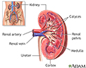
Kidney anatomy - illustration
The kidneys are responsible for removing wastes from the body, regulating electrolyte balance and blood pressure, and stimulating red blood cell production.
Kidney anatomy
illustration
-
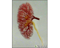
Kidney - blood and urine flow - illustration
This is the typical appearance of the blood vessels (vasculature) and urine flow pattern in the kidney. The blood vessels are shown in red and the urine flow pattern in yellow.
Kidney - blood and urine flow
illustration
-
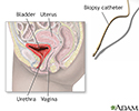
Ureteral biopsy - illustration
The cystoscope enters through the urethra, then the bladder, in order for the guidewire to gain access to the ureter.
Ureteral biopsy
illustration
-
Kidney anatomy - illustration
The kidneys are responsible for removing wastes from the body, regulating electrolyte balance and blood pressure, and stimulating red blood cell production.
Kidney anatomy
illustration
-
Kidney - blood and urine flow - illustration
This is the typical appearance of the blood vessels (vasculature) and urine flow pattern in the kidney. The blood vessels are shown in red and the urine flow pattern in yellow.
Kidney - blood and urine flow
illustration
-
Ureteral biopsy - illustration
The cystoscope enters through the urethra, then the bladder, in order for the guidewire to gain access to the ureter.
Ureteral biopsy
illustration
Review Date: 5/23/2016
Reviewed By: Jennifer Sobol, DO, urologist with the Michigan Institute of Urology, West Bloomfield, MI. Review provided by VeriMed Healthcare Network. Also reviewed by David Zieve, MD, MHA, Isla Ogilvie, PhD, and the A.D.A.M. Editorial team.


