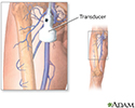
Doppler ultrasound exam of an arm or leg
Peripheral vascular disease - Doppler; PVD - Doppler; PAD - Doppler; Blockage of leg arteries - Doppler; Intermittent claudication - Doppler; Arterial insufficiency of the legs - Doppler; Leg pain and cramping - Doppler; Calf pain - Doppler
This test uses ultrasound to look at the blood flow in the large arteries and veins in the arms and legs.
Ultrasound
Ultrasound uses high-frequency sound waves to make images of organs and structures inside the body.

How the Test is Performed
The test is done in the ultrasound or radiology department or in a peripheral vascular lab.
During the exam:
- A water-soluble gel is placed on a handheld device called a transducer. This device directs high-frequency sound waves to the artery or veins being tested.
- Blood pressure cuffs may be put around different parts of the body, including the thigh, calf, ankle, and different points along the arm.
- A paste is applied to the skin over the arteries being examined. Images are created as the transducer is moved over each area.
How to Prepare for the Test
You will need to remove clothes from the arm or leg being examined.
How the Test will Feel
Sometimes the person performing the test will need to press on the vein to make sure it does not have a clot. Some people may feel slight pain.
Why the Test is Performed
This test is done as the first step to look at arteries and veins. Sometimes arteriography and venography may be needed later. The test is done to help diagnose:
Arteriography
Extremity angiography is a test used to see the arteries in the hands, arms, feet, or legs. It is also called peripheral angiography. Angiography u...
Venography
Venography for legs is a test used to see the veins in the leg. X-rays are a form of electromagnetic radiation, like visible light is. However, thes...

-
Arteriosclerosis of the arms or legs
Arteriosclerosis of the arms or legs
Peripheral artery disease (PAD) is a condition of the blood vessels that supply the legs and feet. It leads to narrowing and hardening of the arteri...
 ImageRead Article Now Book Mark Article
 -
Blood clot
(deep vein thrombosis)
Blood clot
Blood clots are clumps that occur when blood hardens from a liquid to a solid. A blood clot that forms inside one of your veins or arteries is calle...
 ImageRead Article Now Book Mark Article
 -
Venous insufficiency
Venous insufficiency
Venous insufficiency is a condition in which the veins have problems sending blood from the legs back to the heart.
 ImageRead Article Now Book Mark Article
The test may also be used to:
- Look at injury to the arteries
- Monitor arterial reconstruction and bypass grafts
Normal Results
A normal result means the blood vessels show no signs of narrowing, clots, or closure, and the arteries have normal blood flow.
What Abnormal Results Mean
Abnormal results may be due to:
- Blockage in an artery by a blood clot
- Blood clot in a vein (DVT)
- Narrowing or widening of an artery
- Spastic arterial disease (arterial contractions brought on by cold or emotion)
- Venous occlusion (closing of a vein)
- Venous reflux (blood flow going the wrong direction in veins)
- Arterial occlusion from atherosclerosis
This test may also be done to help assess the following conditions:
-
Arteriosclerosis of the extremities
Arteriosclerosis of the extremities
Peripheral artery disease (PAD) is a condition of the blood vessels that supply the legs and feet. It leads to narrowing and hardening of the arteri...
 ImageRead Article Now Book Mark Article
 -
Deep venous thrombosis
Deep venous thrombosis
Deep vein thrombosis (DVT) is a condition that occurs when a blood clot forms in a vein deep inside a part of the body. It mainly affects the large ...
 ImageRead Article Now Book Mark Article
 -
Superficial thrombophlebitis
Superficial thrombophlebitis
Thrombophlebitis is a swollen or inflamed vein due to a blood clot. Superficial refers to veins just below the skin's surface.
 ImageRead Article Now Book Mark Article
Risks
There are no risks from this procedure.
Considerations
Cigarette smoking may alter the results of this test. Nicotine can cause the arteries in the extremities to constrict.
Quitting smoking lowers the risk of problems with the heart and circulatory system. Most smoking-related deaths are caused by cardiovascular problems, not lung cancer.
References
Cosgrove DO, Eckersley RJ, Harvey CJ, Lim A. Ultrasound. In: Adam A, Dixon AK, Gillard JH, Schaefer-Prokop CM, eds. Grainger & Allison's Diagnostic Radiology: A Textbook of Medical Imaging . 6th ed. New York, NY: Elsevier Churchill Livingstone; 2015:chap 3.
Creager MA, Libby P. Peripheral artery diseases. Braunwald's Heart Disease: A Textbook of Cardiovascular Medicine . 10th ed. Philadelphia, PA: Elsevier Saunders; 2015:chap 58.
Rooke TW, Hirsch AT, Misra S, et al. Management of patients with peripheral artery disease (compilation of 2005 and 2011 ACCF/AHA Guideline Recommendations): a report of the American College of Cardiology Foundation/American Heart Association Task Force on Practice Guidelines. J Am Coll Cardiol . 2013;61(14):1555-1570. PMID: 23473760 www.ncbi.nlm.nih.gov/pubmed/23473760 .
-
Doppler ultrasonography of an extremity - illustration
Doppler ultrasonography examines the blood flow in the major arteries and veins in the arms and legs with the use of ultrasound (high-frequency sound waves that echo off the body). It may help diagnose a blood clot, venous insufficiency, arterial occlusion (closing), abnormalities in arterial blood flow caused by a narrowing, or trauma to the arteries.
Doppler ultrasonography of an extremity
illustration
Review Date: 4/2/2015
Reviewed By: Jason Levy, MD, Northside Radiology Associates, Atlanta, GA. Internal review and update on 07/24/2016 by David Zieve, MD, MHA, Isla Ogilvie, PhD, and the A.D.A.M. Editorial team.

