Small intestine aspirate and culture
Small intestine aspirate and culture is a lab test to check for infection in the small intestine.
How the Test is Performed
A sample of fluid from the small intestine is needed. A procedure called an esophagogastroduodenoscopy (EGD ) is done to get the sample.
Esophagogastroduodenoscopy (EGD
Esophagogastroduodenoscopy (EGD) is a test to examine the lining of the esophagus, stomach, and first part of the small intestine.

The fluid is placed in a special dish in the laboratory. It is watched for growth of bacteria or other organisms. This is called a culture.
How the Test will Feel
You are not involved in the test once the sample is taken.
Why the Test is Performed
Your health care provider may order this test if you have signs of too much bacteria growing in the intestinal tract. In most cases, other tests are done first.
Normally, small amounts of bacteria are present in the small intestine and they do not cause disease. However, the test may be done when your doctor suspects that excess growth of intestinal bacteria is causing diarrhea.
Normal Results
No bacteria should be found.
Normal value ranges may vary slightly among different laboratories. Talk to your provider about the meaning of your specific test results.
What Abnormal Results Mean
Abnormal results may be a sign of infection.
Risks
There are no risks associated with a laboratory culture.
References
Beavis KG, Charnot-Katsikas A. Specimen collection and handling for diagnosis of infectious diseases. In: McPherson RA, Pincus MR, eds. Henry's Clinical Diagnosis and Management by Laboratory Methods . 23rd ed. Philadelphia, PA: Elsevier; 2017:chap 64.
Dupont HL. Approach to the patient with suspected enteric infection. In: Goldman L, Schafer AI, eds. Goldman's Cecil Medicine . 25th ed. Philadelphia, PA: Elsevier Saunders; 2016:chap 283.
Fritsche TR, Pritt BS. Medical parasitology. In: McPherson RA, Pincus MR, eds. Henry's Clinical Diagnosis and Management by Laboratory Methods . 23rd ed. Philadelphia, PA: Elsevier; 2017:chap 63.
Gerding DN, Young VB. Clostridium difficle infection. In: Bennett JE, Dolin R, Blaser MJ, eds. Mandell, Douglas, and Bennett's Principles and Practice of Infectious Diseases . 8th ed. Philadelphia, PA: Elsevier Saunders; 2015:chap 245.
Gerding DN, Johnson S. Clostridial infections. In: Goldman L, Schafer AI, eds. Goldman's Cecil Medicine . 25th ed. Philadelphia, PA: Elsevier Saunders; 2016:chap 296.
Haines CF, Sears CL. Infectious enteritis and proctocolitis. In: Feldman M, Friedman LS, Brandt LJ, eds. Sleisenger and Fordtran's Gastrointestinal and Liver Disease . 10th ed. Philadelphia, PA: Elsevier Saunders; 2016:chap 110.
Semrad CE. Approach to the patient with diarrhea and malabsorption. In: Goldman L, Schafer AI, eds. Goldman's Cecil Medicine . 25th ed. Philadelphia, PA: Elsevier Saunders; 2016:chap 140.
Siddiqi HA, Salwen MJ, Shaikh MF, Bowne WB. Laboratory diagnosis of gastrointestinal and pancreatic disorders In: McPherson RA, Pincus MR, eds. Henry's Clinical Diagnosis and Management by Laboratory Methods . 23rd ed. Philadelphia, PA: Elsevier; 2017:chap 22.
-
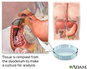
Duodenal tissue culture - illustration
A duodenal tissue biopsy is performed by inserting a special tube through the nose or mouth down into the duodenum. When the tube is in place, it suctions out some of the fluid located in the duodenum. When the procedure is over the tube is removed. The sample is sent to the laboratory for testing. The test is performed to see if a bacterial infection is present or if there are any other microoganisms present that could be causing infection.
Duodenal tissue culture
illustration
Review Date: 5/11/2016
Reviewed By: Subodh K. Lal, MD, gastroenterologist with Gastrointestinal Specialists of Georgia, Austell, GA. Review provided by VeriMed Healthcare Network. Also reviewed by David Zieve, MD, MHA, Isla Ogilvie, PhD, and the A.D.A.M. Editorial team.

