Histoplasma complement fixation
Histoplasma antibody test
Histoplasma complement fixation is a blood test that checks for infection from a fungus called Histoplasma capsulatum ( H capsulatum ), which causes the disease histoplasmosis .
Histoplasmosis
Histoplasmosis is an infection that occurs from breathing in the spores of the fungus Histoplasma capsulatum.

How the Test is Performed
Blood sample is needed
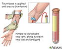
Venipuncture is the collection of blood from a vein. It is most often done for laboratory testing.

The sample is sent to a laboratory. There it is examined for Histoplasma antibodies using a laboratory method called complement fixation. This technique checks if your body has produced substances called antibodies to a specific foreign substance ( antigen ), in this case H capsulatum . Antibodies defend your body against bacteria, viruses, and fungi. If the antibodies are present, they stick, or "fix" themselves, to the antigen. This is why the test is called "fixation."
Complement
Complement is a blood test that measures the activity of certain proteins in the liquid portion of your blood. The complement system is a group of pr...

Antibodies
An antibody is a protein produced by the body's immune system when it detects harmful substances, called antigens. Examples of antigens include micr...

Antigen
An antigen is any substance that causes your immune system to produce antibodies against it. This means your immune system does not recognize the su...

How to Prepare for the Test
There is no special preparation for the test.
How the Test will Feel
When the needle is inserted to draw blood, some people feel moderate pain. Others feel only a prick or stinging. Afterward, there may be some throbbing or bruising. This soon goes away.
Why the Test is Performed
The test is done to detect histoplasmosis infection.
Normal Results
The absence of antibodies (negative test) is normal.
What Abnormal Results Mean
Abnormal results (1:32 or higher) may mean you have an active histoplasmosis infection.
During the early stage of an illness, few antibodies may be detected. Antibody production increases during the course of an infection. For this reason, this test may be repeated several weeks after the first test.
People who have been exposed to H capsulatum in the past may have antibodies to it, often at low levels. But they may not have shown signs of illness.
Risks
Veins and arteries vary in size from one person to another, and from one side of the body to the other. Obtaining a blood sample from some people may be more difficult than from others.
Other risks associated with having blood drawn are slight, but may include:
- Excessive bleeding
- Fainting or feeling lightheaded
- Hematoma (blood accumulating under the skin)
- Infection (a slight risk any time the skin is broken)
References
Deepe GS Jr. Histoplasma capsulatum . In: Bennett JE, Dolin R, Blaser MJ, eds. Mandell, Douglas, and Bennett's Principles and Practice of Infectious Diseases . 8th ed. Philadelphia, PA: Elsevier Saunders; 2015:chap 265.
Iwen PC. Mycotic diseases. In: McPherson RA, Pincus MR, eds. Henry's Clinical Diagnosis and Management by Laboratory Methods . 22nd ed. Philadelphia, PA: Elsevier Saunders; 2011:chap 61.
-
Blood test - illustration
Blood is drawn from a vein (venipuncture), usually from the inside of the elbow or the back of the hand. A needle is inserted into the vein, and the blood is collected in an air-tight vial or a syringe. Preparation may vary depending on the specific test.
Blood test
illustration
Review Date: 5/1/2015
Reviewed By: Jatin M. Vyas, MD, PhD, Assistant Professor in Medicine, Harvard Medical School; Assistant in Medicine, Division of Infectious Disease, Department of Medicine, Massachusetts General Hospital, Boston, MA. Also reviewed by David Zieve, MD, MHA, Bethanne Black, and the A.D.A.M. Editorial Team.

