Extraocular muscle function testing
EOM; Extraocular movement; Ocular motility examination
Extraocular muscle function testing examines the function of the eye muscles. A health care provider observes the movement of the eyes in six specific directions.
How the Test is Performed
You are asked to sit or stand with your head up and looking straight ahead. Your health care provider will hold a pen or other object about 16 inches or 40 centimeters (cm) in front of your face. The provider will then move the object in several directions and ask you to follow it with your eyes, without moving your head.
A test called a cover/uncover test may also be done. You will look at a distant object and the person doing the test will cover the other eye, then after a few seconds, uncover it. You will be asked to keep looking at the distant object. How the eye moves after it is uncovered may show problems. Then the test is performed with the other eye.
How to Prepare for the Test
No special preparation is necessary for this test.
How the Test will Feel
The test involves only normal movement of the eyes.
Why the Test is Performed
This test is performed to evaluate weakness or other problem in the extraocular muscles. These problems may result in double vision or rapid, uncontrolled eye movements .
Rapid, uncontrolled eye movements
Nystagmus is a term to describe fast, uncontrollable movements of the eyes that may be:Side to side (horizontal nystagmus)Up and down (vertical nysta...

Normal Results
Normal movement of the eyes in all directions.
What Abnormal Results Mean
Eye movement disorders may be due to abnormalities of the muscles themselves. They may also be due to problems in the sections of the brain that control these muscles. Your health care provider will talk to you about any abnormalities that may be found.
Risks
There are no risks associated with this test.
Considerations
Rapid uncontrolled eye movements, nystagmus , may occur slightly when shifting the eyes in an extreme sideways direction. This is normal and stops quickly.
Nystagmus
Nystagmus is a term to describe fast, uncontrollable movements of the eyes that may be:Side to side (horizontal nystagmus)Up and down (vertical nysta...

References
American Academy of Ophthalmology Preferred Practice Patterns Committee. Preferred Practice Pattern Guidelines. Comprehensive Adult Medical Eye Evaluation - 2010. Available at one.aao.org/preferred-practice-pattern/comprehensive-adult-medical-eye-evaluation--octobe. Accessed February 27, 2015.
Baloh RW, Jen J. Neuro-ophthalmology. In: Goldman L, Schafer AI, eds. Goldman's Cecil Medicine . 24th ed. Philadelphia, PA: Elsevier Saunders; 2011:chap 432.
Demer JL. Eye movements and positions. In: Tasman W, Jaeger EA, eds. Duane's Ophthalmology . 2013 ed. Philadelphia, PA: Lippincott Williams & Wilkins; 2013:vol 1, chap 2.
Griggs RC, Jozefowicz RF, Aminoff MJ. Approach to the patient with neurologic disease. In: Goldman L, Schafer AI, eds. Goldman's Cecil Medicine . 24th ed. Philadelphia, PA: Elsevier Saunders; 2011:chap 403.
-
Eye - illustration
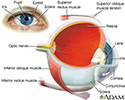
The eye is the organ of sight, a nearly spherical hollow globe filled with fluids (humors). The outer layer or tunic (sclera, or white, and cornea) is fibrous and protective. The middle tunic layer (choroid, ciliary body and the iris) is vascular. The innermost layer (the retina) is nervous or sensory. The fluids in the eye are divided by the lens into the vitreous humor (behind the lens) and the aqueous humor (in front of the lens). The lens itself is flexible and suspended by ligaments which allow it to change shape to focus light on the retina, which is composed of sensory neurons.
Eye
illustration
-
Eye muscle test - illustration
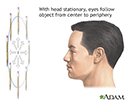
The extraocular muscle function test is performed to evaluate any weakness, or other defect in the extraocular muscles which results in uncontrolled eye movements. The test involves moving the eyes in six different directions in space to evaluate the proper functioning of the extraocular muscles of the eyes.
Eye muscle test
illustration
-
Eye - illustration
The eye is the organ of sight, a nearly spherical hollow globe filled with fluids (humors). The outer layer or tunic (sclera, or white, and cornea) is fibrous and protective. The middle tunic layer (choroid, ciliary body and the iris) is vascular. The innermost layer (the retina) is nervous or sensory. The fluids in the eye are divided by the lens into the vitreous humor (behind the lens) and the aqueous humor (in front of the lens). The lens itself is flexible and suspended by ligaments which allow it to change shape to focus light on the retina, which is composed of sensory neurons.
Eye
illustration
-
Eye muscle test - illustration
The extraocular muscle function test is performed to evaluate any weakness, or other defect in the extraocular muscles which results in uncontrolled eye movements. The test involves moving the eyes in six different directions in space to evaluate the proper functioning of the extraocular muscles of the eyes.
Eye muscle test
illustration
Review Date: 2/23/2015
Reviewed By: Franklin W. Lusby, MD, ophthalmologist, Lusby Vision Institute, La Jolla, CA. Also reviewed by David Zieve, MD, MHA, Isla Ogilvie, PhD, and the A.D.A.M. Editorial team.


