His bundle electrography
His bundle electrogram; HBE; His bundle recording; Electrogram - His bundle; Arrythmia - His; Heart block - His
His bundle electrography is a test that measures electrical activity in a part of the heart that carries the signals that control the time between heartbeats (contractions).
How the Test is Performed
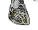
The bundle of His is a group of fibers that carry electrical impulses through the center of the heart. If these signals are blocked, you will have problems with your heartbeat.
The His bundle electrography is part of an electrophysiology (EP) study. An intravenous catheter (IV line) is inserted into your arm so that you can be given medicines during the test.
Electrophysiology
Intracardiac electrophysiology study (EPS) is a test to look at how well the heart's electrical signals are working. It is used to check for abnorma...

Intravenous
Intravenous means "within a vein. " Most often it refers to giving medicines or fluids through a needle or tube inserted into a vein. This allows th...
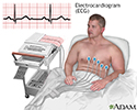
Electrocardiogram (ECG) leads are placed on your arms and legs. Your arm, neck, or groin will be cleaned and numbed with a local anesthetic. After the area is numb, the cardiologist makes a small cut in a vein and inserts a thin tube called a catheter inside.
Electrocardiogram
An electrocardiogram (ECG) is a test that records the electrical activity of the heart.

The catheter is carefully moved through the vein up into the heart. An x-ray method called fluoroscopy helps guide the doctor to the right place. During the test, you are watched for any abnormal heartbeats ( arrhythmias ). The catheter has a sensor on the end, which is used to measure the electrical activity of the bundle of His.
x-ray
X-rays are a type of electromagnetic radiation, just like visible light. An x-ray machine sends individual x-ray particles through the body. The im...

Arrhythmias
An arrhythmia is a disorder of the heart rate (pulse) or heart rhythm. The heart can beat too fast (tachycardia), too slow (bradycardia), or irregul...

How to Prepare for the Test
You will be told not to eat or drink anything for 6 to 8 hours before the test. The test will be done in a hospital. Some people may need to check into the hospital the night before the test. Otherwise, you will check in the morning of the test. Although the test may take some time, most people DO NOT need to stay in the hospital overnight.
Your health care provider will explain the procedure and its risks. You must sign a consent form before the test starts.
About half an hour before the procedure, you will be given a mild sedative to help you relax. You will wear a hospital gown. The procedure may last from 1 to several hours.
How the Test will Feel
You are awake during the test. You may feel some discomfort when the IV is placed into your arm, and some pressure at the site when the catheter is inserted.
Why the Test is Performed
This test may be done to:
- Determine if you need a pacemaker or other treatment
-
Diagnose
arrhythmias
Arrhythmias
An arrhythmia is a disorder of the heart rate (pulse) or heart rhythm. The heart can beat too fast (tachycardia), too slow (bradycardia), or irregul...
 ImageRead Article Now Book Mark Article
 - Find the specific location where electrical signals through the heart are blocked
Normal Results
The time between electrical signals from the bundle of His are evenly spaced.
What Abnormal Results Mean
A pacemaker will be needed if the test results are abnormal.
Abnormal results may mean you have or had:
- Chronic conduction system disease
- Carotid sinus pressure
-
Recent
heart attack
Heart attack
Most heart attacks are caused by a blood clot that blocks one of the coronary arteries. The coronary arteries bring blood and oxygen to the heart. ...
 ImageRead Article Now Book Mark Article
 - Atrial disease
Risks
Risks of the procedure include:
- Arrhythmias
-
Cardiac tamponade
Cardiac tamponade
Cardiac tamponade is pressure on the heart that occurs when blood or fluid builds up in the space between the heart muscle and the outer covering sac...
 ImageRead Article Now Book Mark Article
 -
Embolism from blood clots at the tip of the catheter
Embolism from blood clots at the tip of...
Blood clots are clumps that occur when blood hardens from a liquid to a solid. A blood clot that forms inside one of your veins or arteries is calle...
 ImageRead Article Now Book Mark Article
 -
Heart attack
Heart attack
Most heart attacks are caused by a blood clot that blocks one of the coronary arteries. The coronary arteries bring blood and oxygen to the heart. ...
 ImageRead Article Now Book Mark Article
 - Hemorrhage
- Infection
- Injury to the vein or artery
- Low blood pressure
-
Stroke
Stroke
A stroke occurs when blood flow to a part of the brain stops. A stroke is sometimes called a "brain attack. " If blood flow is cut off for longer th...
 ImageRead Article Now Book Mark Article
References
Chernecky CC, Berger BJ. H. In Chernecky CC, Berger BJ, eds. Laboratory Tests and Diagnostic Procedures . 6th ed. Philadelphia, PA: Elsevier Saunders; 2013:chap H;602-667.
Miller JM, Zipes DP. Diagnosis of cardiac arrhythmias. In: Bonow RO, Mann DL, Zipes DP, Libby P, Braunwald E, eds. Braunwald's Heart Disease: A Textbook of Cardiovascular Medicine . 10th ed. Philadelphia, PA: Elsevier Saunders; 2015:chap 34.
-
Cardiac conduction system
Animation
-
ECG - illustration
The electrocardiogram (ECG, EKG) is used extensively in the diagnosis of heart disease, ranging from congenital heart disease in infants to myocardial infarction and myocarditis in adults. Several different types of electrocardiogram exist.
ECG
illustration
Review Date: 5/5/2016
Reviewed By: Michael A. Chen, MD, PhD, Associate Professor of Medicine, Division of Cardiology, Harborview Medical Center, University of Washington Medical School, Seattle, WA. Also reviewed by David Zieve, MD, MHA, Isla Ogilvie, PhD, and the A.D.A.M. Editorial team.


