Microcephaly
Microcephaly is a condition in which a person's head size is much smaller than that of others of the same age and sex. Head size is measured as the distance around the top of the head. A smaller than normal size is determined using standardized charts.
Causes
Microcephaly most often occurs because the brain does not grow at a normal rate. The growth of the skull is determined by brain growth. Brain growth takes place while a baby is in the womb and during infancy.
Conditions that affect brain growth can cause smaller than normal head size. These include infections, genetic disorders, and severe malnutrition.
Genetic conditions that cause microcephaly include:
- Cornelia de Lange syndrome
-
Cri du chat syndrome
Cri du chat syndrome
Cri du chat syndrome is a group of symptoms that result from missing a piece of chromosome number 5. The syndrome's name is based on the infant's cr...
Read Article Now Book Mark Article - Down syndrome
-
Rubinstein-Taybi syndrome
Rubinstein-Taybi syndrome
Rubinstein-Taybi syndrome (RTS) is a genetic disease. It involves broad thumbs and toes, short stature, distinctive facial features, and varying deg...
 ImageRead Article Now Book Mark Article
 - Seckel syndrome
- Smith-Lemli-Opitz syndrome
-
Trisomy 18
Trisomy 18
Trisomy 18 is a genetic disorder in which a person has a third copy of material from chromosome 18, instead of the usual 2 copies.
 ImageRead Article Now Book Mark Article
 - Trisomy 21
Other problems that may lead to microcephaly include:
- Uncontrolled phenylketonuria (PKU) in the mother
- Methylmercury poisoning
-
Congenital rubella
Congenital rubella
Congenital rubella is a condition that occurs in an infant whose mother is infected with the virus that causes German measles. Congenital means the ...
 ImageRead Article Now Book Mark Article
 -
Congenital toxoplasmosis
Congenital toxoplasmosis
Congenital toxoplasmosis is a group of symptoms that occur when an unborn baby (fetus) is infected with the parasite Toxoplasma gondii.
 ImageRead Article Now Book Mark Article
 -
Congenital cytomegalovirus
(CMV)
Congenital cytomegalovirus
Congenital cytomegalovirus is a condition that can occur when an infant is infected with a virus called cytomegalovirus (CMV) before birth. Congenit...
 ImageRead Article Now Book Mark Article
 - Use of certain drugs during pregnancy, especially alcohol and phenytoin
Becoming infected with the Zika virus while pregnant can also cause microcephaly. The Zika virus is present in Brazil and other parts of South America, along with Mexico, Central America, and the Caribbean.
Zika virus
Zika is a virus passed to humans by the bite of infected mosquitoes. Symptoms include fever, joint pain, rash, and red eyes (conjunctivitis). For th...
When to Contact a Medical Professional
Most often, microcephaly is diagnosed at birth or during routine well-baby exams . Talk to your health care provider if you think your infant's head size is too small or not growing normally.
Well-baby exams
Childhood is a time of rapid growth and change. Children have more well-child visits when they are younger. This is because development is faster d...

Call your health care provider if you or your partner has been to an area where Zika is present and you are pregnant or thinking about becoming pregnant.
What to Expect at Your Office Visit
Most of the time, microcephaly is discovered during a routine exam. Head measurements are part of all well-baby exams for the first 18 months. Tests take only a few seconds while the measuring tape is placed around the infant's head.
The provider will keep a record over time to determine:
- What is the head circumference?
- Is the head growing at a slower rate than the body?
- What other symptoms are there?
It may also be helpful to keep your own records of your baby's growth. Talk to your provider if you notice that the baby's head growth seems to be slowing down.
If your provider diagnoses your child with microcephaly, you should note it in your child's personal medical records.
References
Centers for Disease Control and Prevention. Zika virus. www.cdc.gov/zika/index.html . Accessed February 24, 2016.
Johansson MA, Mier-Y-Teran-Romero L, Reefhuis J, Gilboa SM, Hills SL. Zika and the risk of microcephaly. N Engl J Med . 2016 May 25. PMID: 27222919. www.ncbi.nlm.nih.gov/pubmed/27222919 .
Kinsman SL, Johnston MV. Congenital anomalies of the central nervous system. In: Kliegman RM, Stanton BF, St Geme JW, Schor NF, eds. Nelson Textbook of Pediatrics . 20th ed. Philadelphia, PA: Elsevier; 2016:chap 591.
Mirzaa G, Ashwal S, Dobyns WB. Disorders of brain size. In: Swaiman K, Ashwal S, Ferriero DM, Ferriero D, eds. Swaiman's Pediatric Neurology: Principles and Practice . 5th ed. Philadelphia, PA: Elsevier Saunders; 2012:chap 25.
-
Skull of a newborn - illustration
The "sutures" or anatomical lines where the bony plates of the skull join together can be easily felt in the newborn infant. The diamond shaped space on the top of the skull and the smaller space further to the back are often referred to as the "soft spot" in young infants.
Skull of a newborn
illustration
-
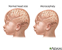
Microcephaly - illustration
Microcephaly is a head size (measured as the distance around the top of the head) significantly below the median for the infant's age and sex. Significantly below is generally considered to be smaller than 3 standard deviations below the mean, or less than 42 cm in circumference at full growth. It most often occurs because of failure of the brain to grow at a normal rate.
Microcephaly
illustration
-
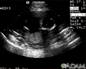
Ultrasound, normal fetus - ventricles of brain - illustration
This is a normal fetal ultrasound performed at 17 weeks gestation. The development of the brain and nervous system begins early in fetal development. During an ultrasound, the technician usually looks for the presence of brain ventricles. Ventricles are spaces in the brain that are filled with fluid. In this early ultrasound, the ventricles can be seen as light lines extending through the skull, seen in the upper right side of the image. The cross hair is pointing to the front of the skull, and directly to the right, the lines of the ventricles are visible.
Ultrasound, normal fetus - ventricles of brain
illustration
-
Skull of a newborn - illustration
The "sutures" or anatomical lines where the bony plates of the skull join together can be easily felt in the newborn infant. The diamond shaped space on the top of the skull and the smaller space further to the back are often referred to as the "soft spot" in young infants.
Skull of a newborn
illustration
-
Microcephaly - illustration
Microcephaly is a head size (measured as the distance around the top of the head) significantly below the median for the infant's age and sex. Significantly below is generally considered to be smaller than 3 standard deviations below the mean, or less than 42 cm in circumference at full growth. It most often occurs because of failure of the brain to grow at a normal rate.
Microcephaly
illustration
-
Ultrasound, normal fetus - ventricles of brain - illustration
This is a normal fetal ultrasound performed at 17 weeks gestation. The development of the brain and nervous system begins early in fetal development. During an ultrasound, the technician usually looks for the presence of brain ventricles. Ventricles are spaces in the brain that are filled with fluid. In this early ultrasound, the ventricles can be seen as light lines extending through the skull, seen in the upper right side of the image. The cross hair is pointing to the front of the skull, and directly to the right, the lines of the ventricles are visible.
Ultrasound, normal fetus - ventricles of brain
illustration
Review Date: 11/19/2015
Reviewed By: Denis Hadjiliadis, MD, MHS, Associate Professor of Medicine, Pulmonary, Allergy, and Critical Care, Perelman School of Medicine, University of Pennsylvania, Philadelphia, PA. Also reviewed by David Zieve, MD, MHA, Isla Ogilvie, PhD, and the A.D.A.M. Editorial team. Editroial update 6/18/2016.


