EEG
Electroencephalogram; Brain wave test; Epilepsy - EEG; Seizure - EEG
An electroencephalogram is a test to measure the electrical activity of the brain.
How the Test is Performed
The test is done by an electroencephalogram (EEG) technologist in your doctor's office or at a hospital or laboratory.
The test is done in the following way:
- You lie on your back on a bed or in a reclining chair.
- Flat metal disks called electrodes are placed all over your scalp. The disks are held in place with a sticky paste. The electrodes are connected by wires to a recording machine. The machine changes the electrical signals into patterns that can be seen on a monitor or drawn on paper. These patterns look like wavy lines.
- You need to lie still during the test with your eyes closed. This is because movement can change the results. You may be asked to do certain things during the test, such as breathe fast and deeply for several minutes or look at a bright flashing light.
- You may be asked to sleep during the test.
If your doctor needs to monitor your brain activity for a longer period, an ambulatory EEG will be ordered. In addition to the electrodes, you will wear or carry a special recorder for up to 3 days. You will be able to go about your normal routine as the EEG is being recorded. Or, your doctor may ask you to stay overnight in a special EEG monitoring unit where your brain activity will be monitored continuously.
How to Prepare for the Test
Wash your hair the night before the test. DO NOT use conditioner, oils, sprays, or gel on your hair. If you have a hair weave, ask your health care provider for special instructions.
Your provider may want you to stop taking certain medicines before the test. DO NOT change or stop taking any medicines without first talking to your provider. Bring a list of your medicines with you.
Avoid all food and drinks containing caffeine for 8 hours before the test.
You may need to sleep during the test. If so, you may be asked to reduce your sleep time the night before. If you are asked to sleep as little as possible before the test, DO NOT eat or drink any caffeine, energy drinks, or other products that help you stay awake.
Follow any other specific instructions you are given.
How the Test will Feel
The electrodes may feel sticky and strange on your scalp, but should not cause any other discomfort. You should not feel any discomfort during the test.
Why the Test is Performed
Brain cells communicate with each other by producing tiny electrical signals, called impulses. An EEG measures this activity. It can be used to diagnose or monitor the following health conditions:
-
Seizures
and
epilepsy
Seizures
A seizure is the physical findings or changes in behavior that occur after an episode of abnormal electrical activity in the brain. The term "seizure...
 ImageRead Article Now Book Mark Article
Epilepsy
Epilepsy is a brain disorder in which a person has repeated seizures over time. Seizures are episodes of uncontrolled and abnormal firing of brain c...
 ImageRead Article Now Book Mark Article
 - Abnormal changes in body chemistry that affect the brain
-
Brain diseases, such as
Alzheimer disease
Alzheimer disease
Dementia is a loss of brain function that occurs with certain diseases. Alzheimer disease is one form of dementia. It affects memory, thinking, and...
 ImageRead Article Now Book Mark Article
 -
Confusion
Confusion
Confusion is the inability to think as clearly or quickly as you normally do. You may feel disoriented and have difficulty paying attention, remembe...
 ImageRead Article Now Book Mark Article
 - Fainting spells or periods of memory loss that cannot be explained otherwise
-
Head injuries
Head injuries
A head injury is any trauma to the scalp, skull, or brain. The injury may be only a minor bump on the skull or a serious brain injury. Head injury c...
 ImageRead Article Now Book Mark Article
 - Infections
- Tumors
EEG is also used to:
-
Evaluate problems with sleep (
sleep disorders
)
Sleep disorders
Sleep disorders are problems with sleeping. These include trouble falling or staying asleep, falling asleep at the wrong times, too much sleep, and ...
 ImageRead Article Now Book Mark Article
 - Monitor the brain during brain surgery
An EEG may be done to show that the brain has no activity, in the case of someone who is in a deep coma. It can be helpful when trying to decide if a person is brain dead.
EEG cannot be used to measure intelligence.
Normal Results
Brain electrical activity has a certain number of waves per second (frequencies) that are normal for different levels of alertness. For example, brain waves are faster when you are awake, and slower in certain stages of sleep.
There are also normal patterns to these waves.
Note: A normal EEG does not mean that a seizure did not occur.
What Abnormal Results Mean
Abnormal results on an EEG test may be due to:
- Abnormal bleeding (hemorrhage)
-
An abnormal structure in the brain (such as a
brain tumor
)
Brain tumor
A brain tumor is a group (mass) of abnormal cells that start in the brain. This article focuses on primary brain tumors in children.
 ImageRead Article Now Book Mark Article
 - Tissue death due to a blockage in blood flow (cerebral infarction)
- Drug or alcohol abuse
- Head injury
- Migraines (in some cases)
- Seizure disorder (such as epilepsy)
-
Sleep disorder (such as
narcolepsy
)
Narcolepsy
Narcolepsy is a nervous system problem that causes extreme sleepiness and attacks of daytime sleep.
 ImageRead Article Now Book Mark Article
 - Swelling of the brain (edema)
Risks
An EEG test is very safe. The flashing lights or fast breathing ( hyperventilation ) required during the test may trigger seizures in those with seizure disorders. The provider performing the EEG is trained to take care of you if this happens.
Hyperventilation
Hyperventilation is rapid and deep breathing. It is also called overbreathing, and it may leave you feeling breathless.
References
Chernecky CC, Berger BJ. Electroencephalography (EEG) - diagnostic. In: Chernecky CC, Berger BJ, eds. Laboratory Tests and Diagnostic Procedures . 6th ed. Philadelphia, PA: Elsevier Saunders; 2013:chap E.
Hahn CD, Emerson RG. Electroencephalography and evoked potentials. In: Daroff RB, Jankovic J, Mazziotta JC, Pomeroy SL, eds. Bradley's Neurology in Clinical Practice . 7th ed. Philadelphia, PA: Elsevier; 2016:chap 34.
-
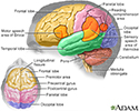
Brain - illustration
The major areas of the brain have one or more specific functions.
Brain
illustration
-
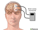
Brain wave monitor - illustration
The brainstem auditory evoked response test (BAER), is performed to help diagnose nervous-system abnormalities, hearing losses (especially in low-birth weight newborns), and to assess neurologic functions. The test focuses on changes and responses in brain waves. The brain waves are stimulated by a clicking sound to evaluate the central auditory pathways of the brainstem.
Brain wave monitor
illustration
-
Brain - illustration
The major areas of the brain have one or more specific functions.
Brain
illustration
-
Brain wave monitor - illustration
The brainstem auditory evoked response test (BAER), is performed to help diagnose nervous-system abnormalities, hearing losses (especially in low-birth weight newborns), and to assess neurologic functions. The test focuses on changes and responses in brain waves. The brain waves are stimulated by a clicking sound to evaluate the central auditory pathways of the brainstem.
Brain wave monitor
illustration
Review Date: 2/27/2016
Reviewed By: Amit M. Shelat, DO, FACP, attending neurologist and Assistant Professor of Clinical Neurology, SUNY Stony Brook, School of Medicine. Review provided by VeriMed Healthcare Network. Also reviewed by David Zieve, MD, MHA, Isla Ogilvie, PhD, and the A.D.A.M. Editorial team.