Center for Cancer Care
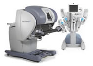
St. Luke's doctors perform minimally-invasive procedures using the da Vinci Surgical System

"I consider the da Vinci System a miracle process, plain and simple. I would recommend it to anyone."
-Roy F., St. Luke's da Vinci Patient
St. Luke's doctors perform minimally-invasive procedures using the da Vinci Surgical System, a robotic and computer-assisted technology that allows our surgeons' hand movements to be scaled, filtered and translated into precise movements within the operative site. The system allows our doctors to gain access to abdominal organs that are affected with better visualization of vital structures using three-dimensional views and enhanced magnification. In addition, the ergonomics of the da Vinci System not only help with physician comfort as they operate, but also assist in aligning their eyes and hands during the procedure. All of these factors help our doctors better identify and avoid sensitive structures such as nerves, veins and arteries, while focusing solely on the surgical site.
St. Luke's is also pleased to introduce the da Vinci® Si™ Surgical System to our operating room and to our community. This is a significant arrival because of the value it offers our surgical staff and those in the region we serve.
The da Vinci® Si™ has several unique features designed to provide additional clinical benefits and efficiency in the operating room, many of which translate to patient benefits. Here are a few features of the da Vinci® Si™:
- Enhanced 3D, high-definition vision of operative field with up to 10x magnification
- New optional dual console allows second surgeon to provide assistance
- Superior visual clarity of tissue and anatomy
- Surgical dexterity and precision far greater than even the human hand
- Updated and simplified user interface to enhance OR efficiency
- New ergonomic settings for greater surgeon comfort
In addition, St. Luke's hospital is the first in the St. Louis area to utilize Firefly, an enhanced fluorescence imaging technology part of the da Vinci® robotic surgical system for surgical removal of part of a kidney.
Fluorescence imaging allows surgeons to see and assess anatomy better than the naked eye further enhancing the unmatched vision, precision and control of minimally invasive da Vinci surgery. Fluorescence imaging, which is an injected dye that glows bright green, helps identify vascular flow to the kidney and distinguish between normal and cancerous tissue. Firefly helps surgeons identify all of the arteries leading to the kidney in order to prevent excessive bleeding during the surgery and preserve the function and blood flow of normal kidney tissue. A specially-designed camera and endoscopes allow the surgeon during the course of the procedure to switch between standard real-time images and images illuminated by the dye.
The injection is performed in three steps. The first injection of the dye by the anesthesiologist helps identify the arteries and areas of blood flow. The second injection of dye helps distinguish normal tissues from tumors because there is less blood flow to tumors. After removal of the tumor, the dye is injected a third time to ensure that the kidney function has fully resumed.
