Aging changes in hormone production
The endocrine system is made up of organs and tissues that produce hormones. Hormones are natural chemicals produced in one location, released into the bloodstream, then used by other target organs and systems.
Endocrine
Endocrine glands release (secrete) hormones into the bloodstream. The endocrine glands include:AdrenalHypothalamusIslets of Langerhans in the pancrea...

Hormones control the target organs. Some organ systems have their own internal control systems along with, or instead of, hormones.
As we age, changes naturally occur in the way body systems are controlled. Some target tissues become less sensitive to their controlling hormone. The amount of hormones produced may also change.
Blood levels of some hormones increase, some decrease, and some are unchanged. Hormones are also broken down (metabolized) more slowly.
Many of the organs that produce hormones are controlled by other hormones. Aging also changes this process. For example, an endocrine tissue may produce less of its hormone than it did at a younger age, or it may produce the same amount at a slower rate.
AGING CHANGES
The hypothalamus is located in the brain. It produces hormones that control the other structures in the endocrine system. The amount of these regulating hormones stays about the same, but the response by the endocrine organs can change as we age.
Hypothalamus
The hypothalamus is an area of the brain that produces hormones that control:Body temperatureHungerMoodRelease of hormones from many glands, especial...
The pituitary gland is also located in the brain. This gland reaches its maximum size in middle age and then gradually becomes smaller. It has two parts:
- The back (posterior) part stores hormones produced in the hypothalamus.
- The front (anterior) part produces hormones that affect growth, the thyroid gland (TSH), adrenal cortex, ovaries, testes, and breasts.
The thyroid gland is located in the neck. It produces hormones that help control metabolism . With aging, the thyroid may become lumpy (nodular). Metabolism slows over time, beginning at around age 20. Because thyroid hormones are produced and broken down (metabolized) at the same rate, thyroid function tests are most often still normal. In some people, thyroid hormone levels may rise, leading to an increased risk of death from cardiovascular disease.
Metabolism
Metabolism refers to all the physical and chemical processes in the body that convert or use energy, such as:BreathingCirculating bloodControlling bo...

Thyroid function tests
Thyroid function tests are used to tell whether your thyroid is working normally. The most common thyroid function tests are:Total, or free T4 (the m...

The parathyroid glands are four tiny glands located around the thyroid. Parathyroid hormone affects calcium and phosphate levels, which affect bone strength. Parathyroid hormone levels rise with age, which may contribute to osteoporosis .
Osteoporosis
Osteoporosis is a disease in which bones become fragile and more likely to break (fracture).

Insulin is produced by the pancreas. It helps sugar (glucose) go from the blood to the inside of cells, where it can be used for energy.
The average fasting glucose level rises 6 to 14 milligrams per deciliter (mg/dL) every 10 years after age 50 as the cells become less sensitive to the effects of insulin.
Fasting glucose
A blood glucose test measures the amount of a sugar called glucose in a sample of your blood. Glucose is a major source of energy for most cells of t...

The adrenal glands are located just above the kidneys. The adrenal cortex, the surface layer, produces the hormones aldosterone , cortisol, and dehydroepiandrosterone.
Adrenal glands
The adrenal glands are two triangle-shaped glands. One gland is located on top of each kidney.
Aldosterone
The aldosterone blood test measures the level of the hormone aldosterone in blood. Aldosterone can also be measured using a urine test.
-
Aldosterone regulates fluid and
electrolyte
balance.
Electrolyte
Electrolytes are minerals in your blood and other body fluids that carry an electric charge. Electrolytes affect how your body functions in many ways...
Read Article Now Book Mark Article - Cortisol is the "stress response" hormone. It affects the breakdown of glucose, protein, and fat, and it has anti-inflammatory and anti-allergy effects.
Aldosterone release decreases with age. This decrease can contribute to lightheadedness and a drop in blood pressure with sudden position changes (orthostatic hypotension). Cortisol release also decreases with aging, but the blood level of this hormone stays about the same. Dehydroepiandrosterone levels also drop. The effects of this drop on the body are not clear.
The ovaries and testes have two functions. They produce the reproductive cells (ova and sperm). They also produce the sex hormones that control secondary sex characteristics, such as breasts and facial hair.
-
With aging, men sometimes have a lower level of
testosterone
.
Testosterone
A testosterone test measures the amount of the male hormone, testosterone, in the blood. Both men and women produce this hormone. The test described...
Read Article Now Book Mark Article -
Women have lower levels of estradiol and other estrogen hormones after
menopause
.
Menopause
Menopause is the time in a woman's life when her periods (menstruation) stop. Most often, it is a natural, normal body change that most often occurs...
 ImageRead Article Now Book Mark Article
EFFECT OF CHANGES
Overall, some hormones decrease, some do not change, and some increase with age. Hormones that usually decrease include:
- Aldosterone
- Calcitonin
-
Growth hormone
Growth hormone
The growth hormone test measures the amount of growth hormone in the blood. The pituitary gland makes growth hormone, which causes a child to grow. ...
 ImageRead Article Now Book Mark Article
 - Renin

In women, estrogen and prolactin levels often decrease significantly.
Prolactin
Prolactin is a hormone released by the pituitary gland. The prolactin test measures the amount of prolactin in the blood.
Hormones that most often remain unchanged or only slightly decrease include:
- Cortisol
- Epinephrine
- Insulin
-
Thyroid hormones
T3
and
T4
T3
Triiodothyronine (T3) is a thyroid hormone. It plays an important role in the body's control of metabolism (the many processes the body does to func...
Read Article Now Book Mark ArticleT4
T4 (thyroxine) is the main hormone produced by the thyroid gland. A laboratory test can be done to measure the amount of free T4 in your blood....
ImageRead Article Now Book Mark Article
Testosterone levels usually decrease gradually as men age.
Hormones that may increase include:
- Follicle-stimulating hormone (FSH)
- Luteinizing hormone (LH)
-
Norepinephrine
Norepinephrine
This test measures the levels of catecholamines in the blood. Catecholamines are hormones made by the adrenal glands. Catecholamines are more often...
ImageRead Article Now Book Mark Article -
Parathyroid hormone
Parathyroid hormone
The PTH test measures the level of parathyroid hormone in the blood. PTH stands for parathyroid hormone. It is a protein hormone released by the par...
Read Article Now Book Mark Article
RELATED TOPICS
-
Aging changes in immunity
Aging changes in immunity
Your immune system helps protect your body from foreign or harmful substances. Examples are bacteria, viruses, toxins, cancer cells, and blood or ti...
 ImageRead Article Now Book Mark Article
 -
Aging changes in organs, tissues, and cells
Aging changes in organs, tissues, and c...
All vital organs begin to lose some function as you age during adulthood. Aging changes occur in all of the body's cells, tissues, and organs, and t...
 ImageRead Article Now Book Mark Article
 -
Aging changes in the male reproductive system
Aging changes in the male reproductive ...
Aging changes in the male reproductive system may include changes in testicular tissue, sperm production, and erectile function. These changes usual...
 ImageRead Article Now Book Mark Article
 -
Menopause
Menopause
Menopause is the time in a woman's life when her periods (menstruation) stop. Most often, it is a natural, normal body change that most often occurs...
ImageRead Article Now Book Mark Article
References
Selmer C, Olesen JB, Hansen ML, et al. Subclinical and overt thyroid dysfunction and risk of all-cause mortality and cardiovascular events: a large population study. J Clin Endocrinol Metab . 2014;99(7):2372-2382. PMID: 24654753 www.ncbi.nlm.nih.gov/pubmed/24654753 .
Skaznik-Wikiel ME, Traub ML, Santoro N. Menopause. In: Jameson JL, De Groot LJ, de Kretser DM, et al, eds. Endocrinology: Adult and Pediatric . 7th ed. Philadelphia, PA: Elsevier Saunders; 2016:chap 135.
Walston JD. Common clinical sequelae of aging. In: Goldman L, Schafer AI, eds. Goldman's Cecil Medicine . 25th ed. Philadelphia, PA: Elsevier Saunders; 2016:chap 25.
-
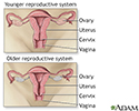
Menopause - illustration
Menopause is the transition in a woman's life when the ovaries stop releasing eggs, menstrual activity decreases and eventually ceases, and the body decreases the production of the female hormones estrogen and progesterone.
Menopause
illustration
-
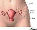
Female reproductive anatomy - illustration
The female reproductive organs are located in the lower abdomen.
Female reproductive anatomy
illustration
-
Menopause - illustration
Menopause is the transition in a woman's life when the ovaries stop releasing eggs, menstrual activity decreases and eventually ceases, and the body decreases the production of the female hormones estrogen and progesterone.
Menopause
illustration
-
Female reproductive anatomy - illustration
The female reproductive organs are located in the lower abdomen.
Female reproductive anatomy
illustration
-
Hypothyroidism
(In-Depth)
-
Menopause
(In-Depth)
-
Dehydroepiandrosterone
(Alt. Medicine)
-
Insomnia
(Alt. Medicine)
-
Osteoporosis
(Alt. Medicine)
-
Menopause
(Alt. Medicine)
-
Prostate cancer
(Alt. Medicine)
-
Melanoma and other skin cancers
(In-Depth)
-
Osteoporosis
(In-Depth)
-
Stress
(In-Depth)
Review Date: 8/22/2016
Reviewed By: Laura J. Martin, MD, MPH, ABIM Board Certified in Internal Medicine and Hospice and Palliative Medicine, Atlanta, GA. Also reviewed by David Zieve, MD, MHA, Isla Ogilvie, PhD, and the A.D.A.M. Editorial team.