Malignant teratoma of the mediastinum
Dermoid cyst - malignant; Nonseminomatous germ cell tumor - teratoma; Immature teratoma; GCTs - teratoma; Teratoma - extragonadal
A teratoma is a type of cancer that contains one or more of the three layers of cells found in a developing baby (embryo). These cells are called germ cells. A teratoma is one type of germ cell tumor.
The mediastinum is located inside the front of the chest in the area that separates the lungs. The heart, large blood vessels, windpipe, thymus gland, and esophagus are found there.
Causes
Malignant mediastinal teratoma occurs most often in young men in their 20s or 30s. Most malignant teratomas can spread throughout the body, and have spread by the time of diagnosis.
Other cancers are often associated with this tumor, including:
-
Acute myelogenous leukemia (AML)
Acute myelogenous leukemia (AML)
Acute myeloid leukemia (AML) is cancer that starts inside bone marrow. This is the soft tissue in the center of bones that helps form all blood cell...
 ImageRead Article Now Book Mark Article
 -
Embryonal rhabdomyosarcoma
Embryonal rhabdomyosarcoma
Rhabdomyosarcoma is a cancerous (malignant) tumor of the muscles that are attached to the bones. This cancer mostly affects children.
 ImageRead Article Now Book Mark Article
 - T-cell lymphoma (a type of blood cancer)
- Myelodysplactic syndromes (group of bone marrow disorders)
- Small cell carcinoma (a type of cancer that grows and spreads rapidly)
Symptoms
Symptoms may include:
- Chest pain or pressure
- Cough
- Fatigue
- Limited ability to tolerate exercise
- Shortness of breath
Exams and Tests
The health care provider will perform a physical exam and ask about the symptoms. The exam may reveal a blockage of the veins entering the center of the chest due to increased pressure in the chest area.
The following tests help diagnose the tumor:
- Chest x-ray
- CT scans of the chest, abdomen, and pelvis
-
Blood tests to check beta-HCG,
alpha fetoprotein
(AFP), and lactate dehydrogenase (LDH) levels
Alpha fetoprotein
Alpha fetoprotein (AFP) is a protein produced by the liver and yolk sac of a developing baby during pregnancy. AFP levels go down soon after birth. ...
 ImageRead Article Now Book Mark Article
 -
Mediastinoscopy with biopsy
Mediastinoscopy with biopsy
Mediastinoscopy with biopsy is a procedure in which a lighted instrument (mediastinoscope) is inserted in the space in the chest between the lungs (m...
 ImageRead Article Now Book Mark Article
Treatment
Chemotherapy is used to treat the tumor. A combination of medicines (usually cisplatin, etoposide, and bleomycin) is commonly used.
Chemotherapy
The term chemotherapy is used to describe cancer-killing drugs. Chemotherapy may be used to:Cure the cancerShrink the cancerPrevent the cancer from ...

After chemotherapy is complete, CT scans are taken again to see if any of the tumor remains. Surgery may be recommended if there is a risk that the cancer will grow back in that area or if any cancer has been left behind.
Support Groups
There are many support groups available for people with cancer. Contact the American Cancer Society -- www.cancer.org
Outlook (Prognosis)
The outlook depends on the tumor size and location and the age of the patient.
Possible Complications
The cancer can spread throughout the body and there may be complications of surgery or related to chemotherapy.
When to Contact a Medical Professional
Call your provider if you have symptoms of malignant teratoma.
References
Cheng G, Varghese TK, Park DR. Mediastinal tumors and cysts. In: Broaddus VC, Mason RJ, Ernst JD, et al, eds. Murray and Nadel's Textbook of Respiratory Medicine . 6th ed. Philadelphia, PA: Elsevier Saunders; 2016:chap 83.
Putnam JB. Lung, chest wall, pleura, and mediastinum. In: Townsend CM, Beauchamp RD, Evers BM Mattox KL, eds. Sabiston Textbook of Surgery . 20th ed. Philadelphia, PA: Elsevier; 2017:chap 57.
-
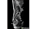
Teratoma - MRI scan - illustration
This MRI scan shows a tumor (teratoma) at the base of the spine (seen on the left lower edge of the screen), located in the sacrum and coccyx (sacrococcygeal) area. Teratomas are present at birth and may contain hair, teeth, and other tissues.
Teratoma - MRI scan
illustration
-
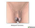
Malignant teratoma - illustration
A malignant teratoma is a type of cancer consisting of cysts that contain one or more of the three primary embryonic germ layers: ectoderm, mesoderm, and endoderm. Because malignant teratomas have usually spread by the time of diagnosis, systemic chemotherapy is needed. The prognosis for people with malignant teratomas is based on the size of the tumor, its location and the age of the patient.
Malignant teratoma
illustration
-
Teratoma - MRI scan - illustration
This MRI scan shows a tumor (teratoma) at the base of the spine (seen on the left lower edge of the screen), located in the sacrum and coccyx (sacrococcygeal) area. Teratomas are present at birth and may contain hair, teeth, and other tissues.
Teratoma - MRI scan
illustration
-
Malignant teratoma - illustration
A malignant teratoma is a type of cancer consisting of cysts that contain one or more of the three primary embryonic germ layers: ectoderm, mesoderm, and endoderm. Because malignant teratomas have usually spread by the time of diagnosis, systemic chemotherapy is needed. The prognosis for people with malignant teratomas is based on the size of the tumor, its location and the age of the patient.
Malignant teratoma
illustration
Review Date: 8/15/2016
Reviewed By: Todd Gersten, MD, Hematology/Oncology, Florida Cancer Specialists & Research Institute, Wellington, FL. Review provided by VeriMed Healthcare Network. Also reviewed by David Zieve, MD, MHA, Isla Ogilvie, PhD, and the A.D.A.M. Editorial team.


