Diskitis
Disk inflammation
Diskitis is swelling (inflammation) and irritation of the space between the bones of the spine (intervertebral disk space).
Causes
Diskitis is an uncommon condition. It is usually seen in children younger than 10 years and in adults around 50 years of age. Men are more affected than women.
Diskitis can be caused by an infection from bacteria or a virus. It can also be caused by inflammation, such as from autoimmune diseases. Autoimmune diseases are conditions in which the immune system mistakenly attacks certain cells in the body.
Disks in the neck and low back are most commonly affected.
Symptoms
Symptoms may include any of the following:
-
Abdominal pain
Abdominal pain
Abdominal pain is pain that you feel anywhere between your chest and groin. This is often referred to as the stomach region or belly.
 ImageRead Article Now Book Mark Article
 - Back pain
- Difficulty getting up and standing
-
Increased
curvature of the back
Curvature of the back
Lordosis is the inward curve of the lumbar spine (just above the buttocks). A small degree of lordosis is normal. Too much curving is called swayba...
 ImageRead Article Now Book Mark Article
 - Irritability
-
Low-grade
fever
(102°F or 38.9°C) or lower
Fever
Fever is the temporary increase in the body's temperature in response to a disease or illness. A child has a fever when the temperature is at or abov...
 ImageRead Article Now Book Mark Article
 -
Sweating at night
- Recent flu-like symptoms
- Refusal to sit up, stand, or walk (younger child)
- Stiffness in back
Exams and Tests
The health care provider will perform a physical exam and ask about the symptoms.
Tests that may be ordered include any of the following:
-
Bone scan
Bone scan
A bone scan is an imaging test used to diagnose bone diseases and find out how severe they are.
 ImageRead Article Now Book Mark Article
 -
Complete blood count (
CBC
)
CBC
A complete blood count (CBC) test measures the following:The number of red blood cells (RBC count)The number of white blood cells (WBC count)The tota...
 ImageRead Article Now Book Mark Article
 -
ESR
or
C-reactive protein
to measure inflammation
ESR
ESR stands for erythrocyte sedimentation rate. It is commonly called a "sed rate. "It is a test that indirectly measures how much inflammation is in...
Read Article Now Book Mark ArticleC-reactive protein
C-reactive protein (CRP) is produced by the liver. The level of CRP rises when there is inflammation throughout the body. It is one of a group of p...
 ImageRead Article Now Book Mark Article
 - MRI of the spine
-
X-ray
of the spine
X-ray
X-rays are a type of electromagnetic radiation, just like visible light. An x-ray machine sends individual x-ray particles through the body. The im...
 ImageRead Article Now Book Mark Article
Treatment
The goal is to treat the cause of the inflammation or infection and reduce pain. Treatment may involve any of the following:
- Antibiotics if the infection is caused by bacteria
- Anti-inflammatory medicines if the cause is an autoimmune disease
- Pain medicines such as NSAIDs
- Bed rest or a brace to keep the back from moving
- Surgery if other methods don't work
Outlook (Prognosis)
Children with an infection should fully recover after treatment. In rare cases, chronic back pain persists.
In cases of autoimmune disease, the outcome depends on the condition. These are often chronic illnesses.
Possible Complications
Complications may include:
- Persistent back pain (rare)
- Side effects of medicines
When to Contact a Medical Professional
Call your provider if your child has back pain that does not go away, or problems with standing and walking that seem unusual for his or her age.
References
Gutierrez K. Diskitis. In: Long SS, ed. Principles and Practice of Pediatric Infectious Diseases . 4th ed. Philadelphia, PA: Elsevier Saunders; 2012:chap 80.
Mistovich RJ, Spiegel DA. The spine. In: Kliegman RM, Stanton BF, St. Geme JW, Schor NF, eds. Nelson Textbook of Pediatrics . 20th ed. Philadelphia, PA: Elsevier; 2016:chap 679.
Waldman SD. Diskitis. In: Waldman SD, ed. Atlas of Common Pain Syndromes . 3rd ed. Philadelphia, PA: Elsevier Saunders; 2012:chap 80.
-
Skeletal spine - illustration
The spine is divided into several sections. The cervical vertebrae make up the neck. The thoracic vertebrae comprise the chest section and have ribs attached. The lumbar vertebrae are the remaining vertebrae below the last thoracic bone and the top of the sacrum. The sacral vertebrae are caged within the bones of the pelvis, and the coccyx represents the terminal vertebrae or vestigial tail.
Skeletal spine
illustration
-
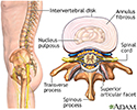
Intervertebral disk - illustration
The vertebral column is made up of 26 bones that provide axial support to the trunk. The vertebral column provides protection to the spinal cord that runs through its central cavity. Between each vertebra is an intervertebral disk. The disks are filled with a gelatinous substance, called the nucleus pulposus, which provides cushioning to the spinal column. The annulus fibrosus is a fibrocartilaginous ring that surrounds the nucleus pulposus, which keeps the nucleus pulposus in tact when forces are applied to the spinal column. The intervertebral disks allow the vertebral column to be flexible and act as shock absorbers during everyday activities such as walking, running and jumping.
Intervertebral disk
illustration
Review Date: 9/22/2016


