Hypersensitivity vasculitis
Allergic vasculitis; Leukocytoclastic vasculitis; Cutaneous vasculitis
Hypersensitivity vasculitis is an extreme reaction to a drug, infection, or foreign substance. It leads to inflammation and damage to blood vessels, primarily in the skin.
Causes
Hypersensitivity vasculitis is caused by an allergic reaction to a drug or other foreign substance. It can also be a reaction to an infection. Most often it affects people older than age 15.
Allergic reaction
Allergic reactions are sensitivities to substances called allergens that come into contact with the skin, nose, eyes, respiratory tract, and gastroin...

Often, the cause of the problem cannot be found even with a careful study of medical history.
Hypersensitivity vasculitis may look like necrotizing vasculitis , which can affect blood vessels throughout the body.
Necrotizing vasculitis
Necrotizing vasculitis is a group of disorders that involve inflammation of the blood vessel walls. The size of the affected blood vessels helps to ...

Symptoms
Symptoms may include:
- New rash over large areas
- Purple-colored spots and patches on the skin
- Skin sores mostly located on the legs, buttocks, or trunk
-
Blisters
on the skin
Blisters
A vesicle is a small fluid-filled blister on the skin.
 ImageRead Article Now Book Mark Article
 -
Hives (
urticaria
), may last longer than 24 hours
Urticaria
Hives are raised, often itchy, red bumps (welts) on the surface of the skin. They are usually an allergic reaction to food or medicine. They can al...
 ImageRead Article Now Book Mark Article
 -
Open sores with dead tissue (necrotic
ulcers
)
Ulcers
An ulcer is a crater-like sore on the skin or mucous membrane. Ulcers form when the top layers of skin or tissue have been removed. They can occur ...
 ImageRead Article Now Book Mark Article
Exams and Tests
The health care provider will base the diagnosis on symptoms. The provider will evaluate how your skin looks after you have taken certain medicines or are exposed to a foreign substance ( antigen ).
Antigen
An antigen is any substance that causes your immune system to produce antibodies against it. This means your immune system does not recognize the su...

Results from an ESR test (erythrocyte sedimentation rate test) may be high.
ESR test
ESR stands for erythrocyte sedimentation rate. It is commonly called a "sed rate. "It is a test that indirectly measures how much inflammation is in...
Skin biopsy shows inflammation of the small blood vessels. You may also have other tests to detect this condition.
Skin biopsy
A skin lesion biopsy is when a small amount of skin is removed so it can be examined. The skin is tested to look for skin conditions or diseases. A...

Treatment
The goal of treatment is to reduce inflammation.
Your provider may prescribe aspirin, nonsteroidal anti-inflammatory drugs (NSAIDs), or corticosteroids to reduce inflammation of the blood vessels. (DO NOT give aspirin to children except as advised by your provider).
Your provider will tell you to stop taking medicines that could be causing this condition.
Outlook (Prognosis)
Hypersensitivity vasculitis most often goes away over time. The condition may come back in some people.
People with ongoing vasculitis should be checked for necrotizing vasculitis.
Possible Complications
Complications may include:
- Lasting damage to the blood vessels or skin with scarring
- Inflamed blood vessels affecting the internal organs
When to Contact a Medical Professional
Call your provider if you have symptoms of hypersensitivity vasculitis.
Prevention
DO NOT take medicines which have caused an allergic reaction in the past.
References
Stone JH. The systemic vasculitides. In: Goldman L, Schafer AI, eds. Goldman's Cecil Medicine . 25th ed. Philadelphia, PA: Elsevier Saunders; 2016:chap 270.
Stone JH. Immune complex-mediated small vessel vasculitis. In: Firestein GS, Budd RC, Gabriel SE, McInnes IB, O'Dell JR, eds. Kelley's Textbook of Rheumatology . 9th ed. Philadelphia, PA: Elsevier Saunders; 2012:chap 91.
-
Vasculitis on the palm - illustration
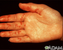
These spots of blood under the skin (purpura) are caused by vasculitis. They do not turn white with pressure (non-blanchable). In this particular case, the purpura are associated with an underlying disorder affecting the structure of the blood vessel walls (collagen-vascular disorder).
Vasculitis on the palm
illustration
-
Vasculitis - illustration
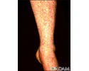
Inflammation of the blood vessels (vasculitis) may be caused when antibodies that have attached to antigens in the blood (immune complexes), attach to the blood vessel walls. These purplish spots can be felt in the skin. They do not turn white (blanch) when pressed. As the condition progresses, they may become larger and more bruise-like (ecchymotic), and some may develop central ulceration or necrosis (tissue death).
Vasculitis
illustration
-
Vasculitis, urticarial on the hand - illustration
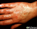
These red (erythematous), hive-like (urticarial) spots (plaques) are caused by inflammation of the blood vessels (urticarial vasculitis) and do not change over a 24-hour period. They may or my not turn white (blanch) with pressure.
Vasculitis, urticarial on the hand
illustration
-
Vasculitis on the palm - illustration
These spots of blood under the skin (purpura) are caused by vasculitis. They do not turn white with pressure (non-blanchable). In this particular case, the purpura are associated with an underlying disorder affecting the structure of the blood vessel walls (collagen-vascular disorder).
Vasculitis on the palm
illustration
-
Vasculitis - illustration
Inflammation of the blood vessels (vasculitis) may be caused when antibodies that have attached to antigens in the blood (immune complexes), attach to the blood vessel walls. These purplish spots can be felt in the skin. They do not turn white (blanch) when pressed. As the condition progresses, they may become larger and more bruise-like (ecchymotic), and some may develop central ulceration or necrosis (tissue death).
Vasculitis
illustration
-
Vasculitis, urticarial on the hand - illustration
These red (erythematous), hive-like (urticarial) spots (plaques) are caused by inflammation of the blood vessels (urticarial vasculitis) and do not change over a 24-hour period. They may or my not turn white (blanch) with pressure.
Vasculitis, urticarial on the hand
illustration
Review Date: 4/28/2015
Reviewed By: Gordon A. Starkebaum, MD, Professor of Medicine, Division of Rheumatology, University of Washington School of Medicine, Seattle, WA. Also reviewed by David Zieve, MD, MHA, Isla Ogilvie, PhD, and the A.D.A.M. Editorial team.


