Eyelid twitch
Eyelid spasm; Eye twitch; Twitch - eyelid; Blepharospasm; Myokymia
An eyelid twitch is a general term for spasms of the eyelid muscles. These spasms happen without your control. The eyelid may repeatedly close (or nearly close) and reopen. This article discusses eyelid twitches in general.
Causes
The most common things that make the muscle in your eyelid twitch are fatigue, stress, and caffeine. Once spasms begin, they may continue off and on for a few days. Then, they disappear. Most people have this type of eyelid twitch once in a while and find it very annoying. In most cases, you won't even notice when the twitch has stopped.
You may have more severe contractions, where the eyelid completely closes. These can be caused by irritation of the:
- Surface of the eye (cornea)
- Membranes lining the eyelids (conjunctiva)
Sometimes, the reason your eyelid is twitching cannot be found. This form of eyelid twitching, called blepharospasm. It lasts much longer and is often very uncomfortable. It can also cause your eyelids to close completely.
Symptoms
Common symptoms of twitching of eyelid are:
- Repeated uncontrollable twitching or spasms of your eyelid (most often the upper lid)
- Light sensitivity (sometimes, this is the cause of the twitching)
- Blurry vision (sometimes)
Treatment
Eyelid twitching most often goes away without treatment. In the meantime, the following steps may help:
- Get more sleep.
- Drink less caffeine.
- Lubricate your eyes with eye drops.
If twitching is severe or lasts a long time, small injections of botulinum toxin can control the spasms.
Outlook (Prognosis)
The outlook depends on the specific type or cause of eyelid twitch. In most cases, the twitches stop within a week.
Possible Complications
Unrecognized cornea injury can cause permanent eye injury. This occurs rarely.
When to Contact a Medical Professional
Call your primary care doctor or eye doctor (ophthalmologist or optometrist) if:
- Eyelid twitching does not go away within 1 week
- Twitching completely closes your eyelid
- Twitching involves other parts of your face
- You have redness, swelling, or a discharge from your eye
-
Your
upper eyelid is drooping
Upper eyelid is drooping
Eyelid drooping is excess sagging of the upper eylid. The edge of the upper eyelid may be lower than it should be (ptosis) or there may be excess ba...
 ImageRead Article Now Book Mark Article
References
Faucett DC. Essential blepharospasm. In: Yanoff M, Duker JS, eds. Ophthalmology . 4th ed. Philadelphia, PA: Elsevier Saunders; 2014:chap 12.8.
Lee AG, Burkat CN, Belinsky I, Marcet MM, Kedar S. Blepharospasm. American Academy of Ophthalmology Web site. Updated March 3, 2016. eyewiki.aao.org/Blepharospasm . Accessed August 31, 2016.
Yanoff M, Cameron JD. Diseases of the visual system. In: Goldman L, Schafer AI, eds. Goldman-Cecil Medicine . 25th ed. Philadelphia, PA: Elsevier Saunders; 2016:chap 423.
-
Eye - illustration
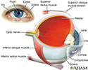
The eye is the organ of sight, a nearly spherical hollow globe filled with fluids (humors). The outer layer or tunic (sclera, or white, and cornea) is fibrous and protective. The middle tunic layer (choroid, ciliary body and the iris) is vascular. The innermost layer (the retina) is nervous or sensory. The fluids in the eye are divided by the lens into the vitreous humor (behind the lens) and the aqueous humor (in front of the lens). The lens itself is flexible and suspended by ligaments which allow it to change shape to focus light on the retina, which is composed of sensory neurons.
Eye
illustration
-
Eye muscles - illustration
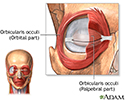
The orbicularis oculi muscles circle the eyes and are located just under the skin. Parts of this muscle act to open and close the eyelids and are important muscles in facial expression.
Eye muscles
illustration
-
Eye - illustration
The eye is the organ of sight, a nearly spherical hollow globe filled with fluids (humors). The outer layer or tunic (sclera, or white, and cornea) is fibrous and protective. The middle tunic layer (choroid, ciliary body and the iris) is vascular. The innermost layer (the retina) is nervous or sensory. The fluids in the eye are divided by the lens into the vitreous humor (behind the lens) and the aqueous humor (in front of the lens). The lens itself is flexible and suspended by ligaments which allow it to change shape to focus light on the retina, which is composed of sensory neurons.
Eye
illustration
-
Eye muscles - illustration
The orbicularis oculi muscles circle the eyes and are located just under the skin. Parts of this muscle act to open and close the eyelids and are important muscles in facial expression.
Eye muscles
illustration
Review Date: 8/20/2016
Reviewed By: Franklin W. Lusby, MD, ophthalmologist, Lusby Vision Institute, La Jolla, CA. Also reviewed by David Zieve, MD, MHA, Isla Ogilvie, PhD, and the A.D.A.M. Editorial team.


