Retroperitoneal fibrosis
Idiopathic retroperitoneal fibrosis; Ormond's disease
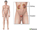
Retroperitoneal fibrosis is a rare disorder that blocks the tubes (ureters) that carry urine from the kidneys to the bladder.
Causes
Retroperitoneal fibrosis occurs when extra fibrous tissue forms in the area behind the stomach and intestines. The tissue forms a mass (or masses) that can block the tubes that carry urine from the kidney to the bladder.
The cause of this problem is not known. It is most common in people aged 40 to 60. Men are twice as likely to develop the condition as women.
Symptoms
Early symptoms:
- Dull pain in the abdomen that increases with time
- Pain and change of color in the legs (due to decreased blood flow)
- Swelling of one leg
Later symptoms:
-
Decreased urine output
Decreased urine output
Decreased urine output means that you produce less urine than normal. Most adults make at least 500 ml of urine in 24 hours (a little over 2 cups)....
 ImageRead Article Now Book Mark Article
 - No urine output (anuria)
- Nausea, vomiting, changes in mental status caused by kidney failure and build-up of toxic chemicals in the blood
- Severe abdominal pain with hemorrhaging (due to death of intestinal tissue)
Exams and tests
Abdominal CT scan is the best way to find a retroperitoneal mass.
Abdominal CT scan
An abdominal CT scan is an imaging method. This test uses x-rays to create cross-sectional pictures of the belly area. CT stands for computed tomog...

Other tests that can help diagnose this condition include:
- BUN and creatinine blood tests
-
Intravenous pyelogram
(IVP), not as commonly used
Intravenous pyelogram
An intravenous pyelogram (IVP) is a special x-ray exam of the kidneys, bladder, and ureters (the tubes that carry urine from the kidneys to the bladd...
 ImageRead Article Now Book Mark Article
 -
Kidney ultrasound
Kidney ultrasound
Abdominal ultrasound is a type of imaging test. It is used to look at organs in the abdomen, including the liver, gallbladder, spleen, pancreas, and...
 ImageRead Article Now Book Mark Article
 - MRI of the abdomen
- CAT scan of the abdomen and retroperitoneum
A biopsy of the mass may also be done to rule out cancer.
Treatment
Corticosteroids are tried first. Some doctors also prescribe a drug called tamoxifen.
If corticosteroid treatment does not work, a biopsy should be done to confirm the diagnosis. Other medicines to suppress the immune system can be prescribed.
When medicine does not work, surgery and stents (draining tubes) are needed.
Stents
A stent is a tiny tube placed into a hollow structure in your body. This structure can be an artery, a blood vessel, or something such as the tube t...

Outlook (Prognosis)
The outlook will depend on the extent of the problem and the amount of damage to the kidneys.
The kidney damage may be temporary or permanent.
Kidney damage
Injury to the kidney and ureter is damage to the organs of the upper urinary tract.

Possible Complications
The disorder may lead to:
- Ongoing blockage of the tubes leading from the kidney on one or both sides
-
Chronic kidney failure
Chronic kidney failure
Chronic kidney disease is the slow loss of kidney function over time. The main job of the kidneys is to remove wastes and excess water from the body...
 ImageRead Article Now Book Mark Article
When to Contact a Medical Professional
Call your health care provider if you have lower abdomen or flank pain and less output of urine.
Prevention
Try to avoid long-term use of medicines that contain methysergide. This drug has been shown to cause retroperitoneal fibrosis. Methysergide is sometimes used to treat migraine headaches.
References
Hsu THS, Nakada SY. Management of upper urinary tract obstruction. In: Wein AJ, ed. Campbell-Walsh Urology . 10th ed. Philadelphia, PA: Elsevier Saunders; 2011:chap 41.
O'Connor OJ, Maher MM. The urinary tract. In: Adam A, Dixon AK, Gillard JH, Schaefer-Prokop CM, eds. Grainger & Allison's Diagnostic Radiology: A Textbook of Medical Imaging . 6th ed. New York, NY: Churchill Livingstone; 2015:chap 35.
Singh I, Strandhopy JW, Assimos DG. Pathophysiology of urinary tract obstruction. In: Wein AJ, ed. Campbell-Walsh Urology . 10th ed. Philadelphia, PA: Elsevier Saunders; 2011:chap 40.
Turnage RH, Richardson KA, Li BD, McDonald JC. Abdominal wall, umbilicus, peritoneum, mesenteries, omentum, and retroperitoneum. In: Townsend CM Jr, Beauchamp RD, Evers BM, Mattox KL, eds. Sabiston Textbook of Surgery . 19th ed. Philadelphia, PA: Elsevier Saunders; 2012:chap 45.
Review Date: 6/29/2015
Reviewed By: Jennifer Sobol, DO, Urologist with the Michigan Institute of Urology, West Bloomfield, MI. Review provided by VeriMed Healthcare Network. Also reviewed by David Zieve, MD, MHA, Isla Ogilvie, PhD, and the A.D.A.M. Editorial team.

