Temporal arteritis
Arteritis - temporal; Cranial arteritis; Giant cell arteritis
Temporal arteritisis inflammation and damage to the blood vessels that supply blood to the head, neck, upper body and arms. It is also called giant cell arteritis.
Causes
Temporal arteritis affects medium-to-large arteries. It causes inflammation, swelling, tenderness, and damage to the blood vessels that supply blood to the head, neck, upper body and arms. It most commonly occurs in the arteries around the temples (temporal arteries) that branch off from the carotid artery in the neck. In some cases, the condition can occur in medium-to-large arteries in other places in the body as well.
The cause of the condition is unknown. It is believed to be due in part to a faulty immune response . The disorder has been linked to some infections and to certain genes.
Immune response
The immune response is how your body recognizes and defends itself against bacteria, viruses, and substances that appear foreign and harmful....

The problem may develop with or following another inflammatory disorder known as polymyalgia rheumatica . Giant cell arteritis almost always occurs in people over age 50. It is most common in people of northern European descent. The condition may run in families.
Polymyalgia rheumatica
Polymyalgia rheumatica (PMR) is an inflammatory disorder. It involves pain and stiffness in the shoulders and often the hips.
Symptoms
Some common symptoms of this problem are:
- Throbbing headache on one side of the head or the back of the head
- Tenderness when touching the scalp
Other symptoms may include:
- Fever
- General ill feeling
- Jaw pain that occurs when chewing
- Pain in the arm after using it
- Muscle aches
- Pain and stiffness in the neck, upper arms, shoulder, and hips
- Weakness, excessive tiredness
Problems with eyesight may occur, and at times may begin suddenly. These problems include:
- Blurred vision
- Double vision
- Reduced vision (blindness in one or both eyes)
Other symptoms may occur with this disease, including:
- Cough
- Tongue or throat pain
-
Hearing loss
Hearing loss
Hearing loss is being partly or totally unable to hear sound in one or both ears.
 ImageRead Article Now Book Mark Article
 - Joint stiffness
Exams and Tests
The health care provider will examine your head.
- The scalp is often sensitive to touch
- There may be a tender, thick artery on one side of the head, most often over one or both temples.
Blood tests may include:
-
Hemoglobin
or
hematocrit
Hemoglobin
Hemoglobin is a protein in red blood cells that carries oxygen. The hemoglobin test measures how much hemoglobin is in your blood.
 ImageRead Article Now Book Mark Article
Hematocrit
Hematocrit is a blood test that measures how much of a person's blood is made up of red blood cells. This measurement depends on the number of and s...
 ImageRead Article Now Book Mark Article
 -
Liver function tests
Liver function tests
Liver function tests are common tests that are used to see how well the liver is working. Tests include:AlbuminAlpha-1 antitrypsin Alkaline phosph...
 ImageRead Article Now Book Mark Article
 -
Sedimentation rate
(ESR) and
C-reactive protein
Sedimentation rate
ESR stands for erythrocyte sedimentation rate. It is commonly called a "sed rate. "It is a test that indirectly measures how much inflammation is in...
Read Article Now Book Mark ArticleC-reactive protein
C-reactive protein (CRP) is produced by the liver. The level of CRP rises when there is inflammation throughout the body. It is one of a group of p...
 ImageRead Article Now Book Mark Article
Blood tests alone cannot provide a diagnosis. You will need to have a biopsy (tissue sample) from the involved artery.
Biopsy
A biopsy is the removal of a small piece of tissue for laboratory examination.
You may also have other tests, including:
-
Duplex ultrasound
Duplex ultrasound
A duplex ultrasound is a test to see how blood moves through your arteries and veins.
 ImageRead Article Now Book Mark Article
 -
MRI
MRI
A magnetic resonance imaging (MRI) scan is an imaging test that uses powerful magnets and radio waves to create pictures of the body. It does not us...
 ImageRead Article Now Book Mark Article
 -
PET scan
PET scan
A positron emission tomography scan is a type of imaging test. It uses a radioactive substance called a tracer to look for disease in the body. A po...
 ImageRead Article Now Book Mark Article
Treatment
Getting prompt treatment can help prevent severe problems such as blindness or even stroke.
Most of the time, you will receive corticosteroids such as prednisone by mouth. These medicines are often started even before a biopsy is done. You may also be told to take aspirin.
Most people begin to feel better within a few days after starting treatment. However, you will need to take medicine for 1 to 2 years. The dose of corticosteroids will be cut back very slowly.
Long-term treatment with corticosteroids can make bones thinner and increase your chance of a fracture. You will need to take the following steps to protect your bone strength.
- Avoid smoking and excess alcohol intake
- Take extra calcium and vitamin D (based on your health care provider's advice)
- Start walking or other forms of weight-bearing exercises
-
Have your bones checked with a
bone mineral density
(BMD) test or DEXA scan
Bone mineral density
A bone mineral density (BMD) test measures how much calcium and other types of minerals are in an area of your bone. This test helps your health care...
 ImageRead Article Now Book Mark Article
 - Take a bisphosphonate medicine such as alendronate (Fosamax) as prescribed by your provider.
You may also need to take other medications that suppress the immune system.
Outlook (Prognosis)
Most people make a full recovery, but treatment may be needed for 1 to 2 years or longer. The condition may return at a later date.
Damage to other blood vessels in the body such as aneurysms (ballooning of the blood vessels) may occur. This damage can lead to a stroke in the future.
When to Contact a Medical Professional
Call your health care provider if you have:
- Throbbing headache that does not go away
- Loss of vision
- Other symptoms of temporal arteritis
Prevention
There is no known prevention.
References
Hellmann DB. Giant cell arteritis, polymyalgia rheumatica, and Takayasu's arteritis. In: Firestein GS, Budd RC, Gabriel SE, et al, eds. Kelley's Textbook of Rheumatology . 9th ed. Philadelphia, PA: Elsevier Saunders; 2012:chap 88.
-
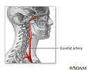
Carotid artery anatomy - illustration
There are four carotid arteries, two on each side of the neck: right and left internal carotid arteries, and right and left external carotid arteries. The carotid arteries deliver oxygen-rich blood from the heart to the head and brain.
Carotid artery anatomy
illustration
Review Date: 1/20/2015
Reviewed By: Gordon A. Starkebaum, MD, professor of medicine, division of rheumatology, University of Washington School of Medicine, Seattle, WA. Also reviewed by David Zieve, MD, MHA, Isla Ogilvie, PhD, and the A.D.A.M. Editorial team.


