Lumbosacral spine CT
Spinal CT; CT - lumbosacral spine; Low back pain - CT; LBP - CT
A lumbosacral spine CT is a computed tomography scan of the lower spine and surrounding tissues.
Computed tomography
A computed tomography (CT) scan is an imaging method that uses x-rays to create pictures of cross-sections of the body. Related tests include:Abdomin...

How the Test is Performed
You will be asked to lie on a narrow table that slides into the center of the CT scanner. You will need to lie on your back for this test.
Once inside the scanner, the machine's x-ray beam rotates around you.
Small detectors inside the scanner measure the amount of x-rays that make it through the part of the body being studied. A computer takes this information and uses it to create a number of images, called slices. These images can be stored, viewed on a monitor, or printed on film. Three-dimensional models of organs can be created by stacking the individual slices together.
You must be still during the exam, because movement causes blurred images. You may be told to hold your breath for short periods.
In some cases, an iodine-based dye, called contrast, may be injected into your vein before images are taken. Contrast can highlight specific areas inside the body, which creates a clearer image.
In other cases, a CT of the lumbosacral spine is done after injecting contrast dye into the spinal canal during a lumbar puncture to further check for pressure on the nerves.
Lumbar puncture
Cerebrospinal fluid (CSF) collection is a test to look at the fluid that surrounds the brain and spinal cord. CSF acts as a cushion, protecting the b...

The scan usually lasts a few minutes.
How to Prepare for the Test
You should remove all jewelry or other metal objects before the test. This is because they may cause inaccurate and blurry images.
How the Test will Feel
The x-rays are painless. Some people may have discomfort from lying on the hard table.
Contrast may cause a slight burning sensation, a metallic taste in the mouth, and a warm flushing of the body. These sensations are normal and usually go away within a few seconds.
Why the Test is Performed
CT rapidly creates detailed pictures of the body. A CT of the lumbosacral spine can evaluate fractures and changes of the spine, such as those due to arthritis or deformities.
What Abnormal Results Mean
CT of the lumbosacral spine may reveal the following conditions or diseases:
-
Cyst
Cyst
A cyst is a closed pocket or pouch of tissue. It can be filled with air, fluid, pus, or other material.
Read Article Now Book Mark Article -
Herniated disk
Herniated disk
A herniated (slipped) disk occurs when all or part of a disk is forced through a weakened part of the disk. This may place pressure on nearby nerves...
 ImageRead Article Now Book Mark Article
 - Infection
- Cancer that has spread to the spine
-
Osteoarthritis
Osteoarthritis
Osteoarthritis (OA) is the most common joint disorder. It is due to aging and wear and tear on a joint.
 ImageRead Article Now Book Mark Article
 -
Osteomalacia
(softening of the bones)
Osteomalacia
Osteomalacia is softening of the bones. It most often occurs because of a problem with vitamin D, which helps your body absorb calcium. Your body n...
 ImageRead Article Now Book Mark Article
 - Pinched nerve
-
Tumor
Tumor
A tumor is an abnormal growth of body tissue. Tumors can be cancerous (malignant) or noncancerous (benign).
Read Article Now Book Mark Article -
Vertebral
fracture
(broken spine bone)
Fracture
If more pressure is put on a bone than it can stand, it will split or break. A break of any size is called a fracture. If the broken bone punctures...
 ImageRead Article Now Book Mark Article
Risks
The most common type of contrast given into a vein contains iodine. If a person with an iodine allergy is given this type of contrast, hives , itching , nausea , breathing difficulty , or other symptoms may occur.
Hives
Hives are raised, often itchy, red bumps (welts) on the surface of the skin. They are usually an allergic reaction to food or medicine. They can al...

Itching
Itching is a tingling or irritation of the skin that makes you want to scratch the area. Itching may occur all over the body or only in one location...

Nausea
Nausea is feeling an urge to vomit. It is often called "being sick to your stomach. "Vomiting or throwing-up is forcing the contents of the stomach ...

Breathing difficulty
Breathing difficulty may involve:Difficult breathingUncomfortable breathingFeeling like you are not getting enough air

If you have kidney problems, diabetes or are on kidney dialysis, talk to your health care provider before the test about your risks.
CT scans and other x-rays are strictly monitored and controlled to make sure they use the least amount of radiation. The risk associated with any individual scan is small. The risk increases when many more scans are performed.
In some cases, a CT scan may still be done if the benefits greatly outweigh the risks. For example, it can be more risky not to have the exam if your provider thinks you might have cancer.
Pregnant or breastfeeding women should consult their provider about the risk of CT scans to the baby. Radiation during pregnancy can affect the baby, and the dye used with CT scans can enter breast milk.
References
Shaw AS, Prokop M. Computed tomography. In: Adam A, Dixon AK, Gillard JH, Schaefer-Prokop CM, eds. Grainger & Allison's Diagnostic Radiology . 6th ed. Philadelphia, PA: Elsevier Churchill Livingstone; 2015:chap 4.
Thomsen HS, Reimer P. Intravascular contrast media for radiography, CT, MRI, and ultrasound. In: Adam A, Dixon AK, Gillard JH, Schaefer-Prokop CM, eds. Grainger & Allison's Diagnostic Radiology . 6th ed. Philadelphia, PA: Elsevier Churchill Livingstone; 2015:chap 2.
-
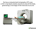
CT scan - illustration
CT stands for computerized tomography. In this procedure, a thin X-ray beam is rotated around the area of the body to be visualized. Using very complicated mathematical processes called algorithms, the computer is able to generate a 3-D image of a section through the body. CT scans are very detailed and provide excellent information for the physician.
CT scan
illustration
-
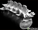
Skeletal spine - illustration
The spine is divided into several sections. The cervical vertebrae make up the neck. The thoracic vertebrae comprise the chest section and have ribs attached. The lumbar vertebrae are the remaining vertebrae below the last thoracic bone and the top of the sacrum. The sacral vertebrae are caged within the bones of the pelvis, and the coccyx represents the terminal vertebrae or vestigial tail.
Skeletal spine
illustration
-
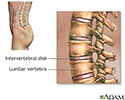
Vertebra, lumbar (low back) - illustration
These are the five vertebra of the lower back. The last vertebra (on the upper left of the picture) attaches to the sacrum, and the top vertebra (on the right of the picture) attaches to the thoracic section of the back. The vertebra are broader and stronger than the other bones in the spine. This allows them to absorb the added pressure applied to the lower back, but this area remains a common site of injury. The vertebra are numbered from one to five and are labeled L1, L2, L3 etc. from the higher bones to the lower.
Vertebra, lumbar (low back)
illustration
-
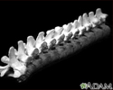
Vertebra, thoracic (mid back) - illustration
These are twelve vertebra of the mid back. The last vertebra (on the left side of the picture) attaches to the lumbar (lower) spine, and the top vertebra (on the right) attaches to the cervical (neck) section of the back. The vertebra are broader and stronger than the cervical bones. This allows them to absorb the added pressure applied to the mid back, but they remain a common sight of injury. The vertebra are numbered from one to twelve and labeled T1, T2, T3, et cetera, from the upper most bones to the lowest.
Vertebra, thoracic (mid back)
illustration
-
Lumbar vertebrae - illustration
There are five lumbar vertebrae located in the lower back. These vertebrae receive the most stress and are the weight-bearing portion of the back. The lumbar vertebrae allow movements such as flexion and extension, and some lateral flexion.
Lumbar vertebrae
illustration

-
CT scan - illustration
CT stands for computerized tomography. In this procedure, a thin X-ray beam is rotated around the area of the body to be visualized. Using very complicated mathematical processes called algorithms, the computer is able to generate a 3-D image of a section through the body. CT scans are very detailed and provide excellent information for the physician.
CT scan
illustration
-
Skeletal spine - illustration
The spine is divided into several sections. The cervical vertebrae make up the neck. The thoracic vertebrae comprise the chest section and have ribs attached. The lumbar vertebrae are the remaining vertebrae below the last thoracic bone and the top of the sacrum. The sacral vertebrae are caged within the bones of the pelvis, and the coccyx represents the terminal vertebrae or vestigial tail.
Skeletal spine
illustration
-
Vertebra, lumbar (low back) - illustration
These are the five vertebra of the lower back. The last vertebra (on the upper left of the picture) attaches to the sacrum, and the top vertebra (on the right of the picture) attaches to the thoracic section of the back. The vertebra are broader and stronger than the other bones in the spine. This allows them to absorb the added pressure applied to the lower back, but this area remains a common site of injury. The vertebra are numbered from one to five and are labeled L1, L2, L3 etc. from the higher bones to the lower.
Vertebra, lumbar (low back)
illustration
-
Vertebra, thoracic (mid back) - illustration
These are twelve vertebra of the mid back. The last vertebra (on the left side of the picture) attaches to the lumbar (lower) spine, and the top vertebra (on the right) attaches to the cervical (neck) section of the back. The vertebra are broader and stronger than the cervical bones. This allows them to absorb the added pressure applied to the mid back, but they remain a common sight of injury. The vertebra are numbered from one to twelve and labeled T1, T2, T3, et cetera, from the upper most bones to the lowest.
Vertebra, thoracic (mid back)
illustration
-
Lumbar vertebrae - illustration
There are five lumbar vertebrae located in the lower back. These vertebrae receive the most stress and are the weight-bearing portion of the back. The lumbar vertebrae allow movements such as flexion and extension, and some lateral flexion.
Lumbar vertebrae
illustration
Review Date: 9/22/2016
Reviewed By: C. Benjamin Ma, MD, Professor, Chief, Sports Medicine and Shoulder Service, UCSF Department of Orthopaedic Surgery, San Francisco, CA. Also reviewed by David Zieve, MD, MHA, Isla Ogilvie, PhD, and the A.D.A.M. Editorial team.