Placenta abruptio
Premature placental separation; Placental separation; Placental abruption; Vaginal bleeding - abruption; Pregnancy - abruption
What is Placenta Abruptio?
The placenta connects the fetus (unborn baby) to the mother's uterus. It allows the baby to get nutrients, blood, and oxygen from the mother. It also helps the baby get rid of waste.
Placenta abruptio (also called placental abruption) is when the placenta separates from the inner wall of the uterus before the baby is born.
More About This Condition
In most pregnancies, the placenta stays attached to the upper part of the uterine wall.
In a small number of pregnancies, the placenta detaches (pulls itself from the wall of the uterus) too early. Most of the time, only part of the placenta pulls away. Other times it pulls away completely. If this happens, it is most often in the 3rd trimester.
The placenta is the lifeline of a fetus. Serious problems occur if it detaches. The baby gets less oxygen and fewer nutrients. Some babies become growth restricted (very small), and in a small number of cases, it is fatal. It can also cause significant blood loss for the mother.
What Causes It?
No one knows what causes placental abruption. But these factors raise a woman's risk for it:
- Long-term (chronic) high blood pressure
- Sudden high blood pressure in pregnant women who had normal blood pressure in the past
- Heart disease
- Diabetes
- Smoking
- Alcohol or cocaine use
- Placental abruption in an earlier pregnancy
- An injury to the mother (such as a car crash or fall in which the abdomen was hit)
- Being African American
- Being older than 40
Signs of Placental Abruption
The most common symptoms are vaginal bleeding and painful contractions. The amount of bleeding depends on how much of the placenta has detached. Sometimes the blood that collects when the placenta detaches stays between the placenta and uterine wall, so you may not have bleeding from your vagina.
- If the separation is slight, you may have only light bleeding. You may also have cramps or feel tender in your belly.
- If the separation is moderate, you may have heavier bleeding. Cramps and belly pain will be more severe.
- If more than half the placenta detaches, you may have belly pain and heavy bleeding. You may also have contractions. The baby may move more or less than normal.
If you have any of these symptoms during your pregnancy, tell your health care provider right away.
How is Placental Abruption Treated?
Your provider will:
- Do a physical exam
- Observe your contractions and how your baby responds to them
- Sometimes do an ultrasound to check your placenta (but ultrasound does not always show a placental abruption)
- Check your baby's heart rate and rhythm
If your placental abruption is small, your provider may put you on bed rest to stop your bleeding. After a few days, most women can go back to their normal activities in most cases.
For a moderate separation, you will likely need to stay in the hospital. In the hospital:
- Your baby's heart rate will be monitored.
- You might need a blood transfusion.
-
If your baby shows any signs of distress, your provider may induce your labor early. If you cannot give birth vaginally, you will need a
C-section
.
C-section
A C-section is the delivery of a baby through a surgical opening in the mother's lower belly area. It is also called a cesarean delivery.
 ImageRead Article Now Book Mark Article
Severe placental abruption is an emergency. You will need to deliver right away, most often by C-section. It is very rare, but a baby can be stillborn if there is a severe abruption.
Can I Prevent Placental Abruption?
You cannot prevent placental abruption, but you can control the risk factors related to it by:
- Keeping high blood pressure, heart disease, and diabetes under control
- Not using tobacco, alcohol, cocaine, or amphetamines
- Following your provider's recommendations about ways to lower your risk if you had an abruption in a past pregnancy
References
Francois KE, Foley MR. Antepartum and postpartum hemorrhage. In: Gabbe SG, Niebyl JR, Simpson JL, et al, eds. Obstetrics: Normal and Problem Pregnancies . 7th ed. Philadelphia, PA: Elsevier; 2017:chap 18.
Hull AD, Resnik R. Placenta previa, placenta accreta, abruptio placentae, and vasa previa. In: Creasy RK, Resnik R, Iams JD, Lockwood CJ, Moore TR, Greene MF, eds. Creasy and Resnik's Maternal-Fetal Medicine: Principles and Practice . 7th ed. Philadelphia, PA: Elsevier Saunders; 2014:chap 46.
-
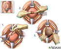
Cesarean section - illustration
The uterus is exposed through the abdominal wall, and an incision is made in the uterine covering. The muscles of the uterus are separated, producing a hole for the delivery of the infant. The infant is delivered through the opening in the uterine wall, after which, the uterus is stitched closed. The uterus is exposed through the abdominal wall, and an incision is made in the uterine covering. The muscles of the uterus are separated, producing a hole for the delivery of the infant. The infant is delivered through the opening in the uterine wall, after which, the uterus is stitched closed.
Cesarean section
illustration
-
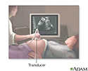
Ultrasound in pregnancy - illustration
The ultrasound has become a standard procedure used during pregnancy. It can demonstrate fetal growth and can detect increasing numbers of conditions in the fetus including meningomyelocele, congenital heart disease, kidney abnormalities, hydrocephalus, anencephaly, club feet, and other deformities. Ultrasound does not produce ionizing radiation and is considered a very safe procedure for both the mother and the fetus.
Ultrasound in pregnancy
illustration
-
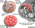
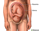
Anatomy of a normal placenta - illustration
The placenta provides the fetus with oxygen and nutrients and takes away waste such as carbon dioxide via the umbilical cord.
Anatomy of a normal placenta
illustration
-
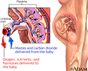
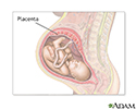
Placenta - illustration
In the placenta, nutrients, wastes, and gases are exchanged between the mother's blood and the baby's blood.
Placenta
illustration
-
Placenta - illustration
The placenta grows during pregnancy and stays connected to the wall of the uterus where it provides the fetus with nourishment. The placenta also secretes hormones that help regulate and maintain the pregnancy.
Placenta
illustration
-
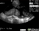
Ultrasound, normal placenta - Braxton Hicks - illustration
This is a normal ultrasound performed at 17 weeks gestation. It shows the placenta during a normal (Braxton Hicks) contraction. Throughout the pregnancy, the uterus periodically contracts to facilitate better blood flow through the placenta and the fetus. In this ultrasound, the placenta can be seen as the mound-shaped object in the middle of the screen. At the bottom of the image, the mother's vertebra can be seen as a round object. When the uterus is not contracting, the placenta would appear much flatter.
Ultrasound, normal placenta - Braxton Hicks
illustration
-
Ultrasound, normal fetus - arms and legs - illustration
This is a normal fetal ultrasound performed at 19 weeks gestation. This is the type of spilt-screen display you might see during an ultrasound, or if the technician prints a copy of the ultrasound for you. This ultrasound shows both the left arm (seen in the left side of the display), and the lower extremities (seen in the right side of the display). The white areas of the arm or legs is developing bone.
Ultrasound, normal fetus - arms and legs
illustration
-
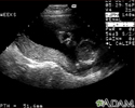
Ultrasound, normal relaxed placenta - illustration
This is a normal fetal ultrasound performed at 19 weeks gestation. This ultrasound shows two interesting features. In the foreground, to the left and middle of the screen, you can see the placenta, following the curve of the uterus. In the background on the right, where the cross hair is pointing, you can see the face with all the facial features visible.
Ultrasound, normal relaxed placenta
illustration
-
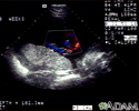
Ultrasound, color - normal umbilical cord - illustration
This is a normal color Doppler ultrasound of the umbilical cord performed at 30 weeks gestation. The cord is the colored area in the middle of the screen, with the different blood vessels represented by different colors. There are normally three vessels in the cord, two arteries and one vein. The umbilical cord is connected to the placenta, located in the middle left of the image.
Ultrasound, color - normal umbilical cord
illustration
-
Placenta - illustration
The placenta supplies the fetus with the blood supply and nutrients necessary for survival.
Placenta
illustration

-
Cesarean section - illustration
The uterus is exposed through the abdominal wall, and an incision is made in the uterine covering. The muscles of the uterus are separated, producing a hole for the delivery of the infant. The infant is delivered through the opening in the uterine wall, after which, the uterus is stitched closed. The uterus is exposed through the abdominal wall, and an incision is made in the uterine covering. The muscles of the uterus are separated, producing a hole for the delivery of the infant. The infant is delivered through the opening in the uterine wall, after which, the uterus is stitched closed.
Cesarean section
illustration
-
Ultrasound in pregnancy - illustration
The ultrasound has become a standard procedure used during pregnancy. It can demonstrate fetal growth and can detect increasing numbers of conditions in the fetus including meningomyelocele, congenital heart disease, kidney abnormalities, hydrocephalus, anencephaly, club feet, and other deformities. Ultrasound does not produce ionizing radiation and is considered a very safe procedure for both the mother and the fetus.
Ultrasound in pregnancy
illustration
-
Anatomy of a normal placenta - illustration
The placenta provides the fetus with oxygen and nutrients and takes away waste such as carbon dioxide via the umbilical cord.
Anatomy of a normal placenta
illustration
-
Placenta - illustration
In the placenta, nutrients, wastes, and gases are exchanged between the mother's blood and the baby's blood.
Placenta
illustration
-
Placenta - illustration
The placenta grows during pregnancy and stays connected to the wall of the uterus where it provides the fetus with nourishment. The placenta also secretes hormones that help regulate and maintain the pregnancy.
Placenta
illustration
-
Ultrasound, normal placenta - Braxton Hicks - illustration
This is a normal ultrasound performed at 17 weeks gestation. It shows the placenta during a normal (Braxton Hicks) contraction. Throughout the pregnancy, the uterus periodically contracts to facilitate better blood flow through the placenta and the fetus. In this ultrasound, the placenta can be seen as the mound-shaped object in the middle of the screen. At the bottom of the image, the mother's vertebra can be seen as a round object. When the uterus is not contracting, the placenta would appear much flatter.
Ultrasound, normal placenta - Braxton Hicks
illustration
-
Ultrasound, normal fetus - arms and legs - illustration
This is a normal fetal ultrasound performed at 19 weeks gestation. This is the type of spilt-screen display you might see during an ultrasound, or if the technician prints a copy of the ultrasound for you. This ultrasound shows both the left arm (seen in the left side of the display), and the lower extremities (seen in the right side of the display). The white areas of the arm or legs is developing bone.
Ultrasound, normal fetus - arms and legs
illustration
-
Ultrasound, normal relaxed placenta - illustration
This is a normal fetal ultrasound performed at 19 weeks gestation. This ultrasound shows two interesting features. In the foreground, to the left and middle of the screen, you can see the placenta, following the curve of the uterus. In the background on the right, where the cross hair is pointing, you can see the face with all the facial features visible.
Ultrasound, normal relaxed placenta
illustration
-
Ultrasound, color - normal umbilical cord - illustration
This is a normal color Doppler ultrasound of the umbilical cord performed at 30 weeks gestation. The cord is the colored area in the middle of the screen, with the different blood vessels represented by different colors. There are normally three vessels in the cord, two arteries and one vein. The umbilical cord is connected to the placenta, located in the middle left of the image.
Ultrasound, color - normal umbilical cord
illustration
-
Placenta - illustration
The placenta supplies the fetus with the blood supply and nutrients necessary for survival.
Placenta
illustration
Review Date: 11/11/2016
Reviewed By: Irina Burd, MD, PhD, Associate Professor of Gynecology and Obstetrics at Johns Hopkins University School of Medicine, Baltimore, MD. Review provided by VeriMed Healthcare Network. Also reviewed by David Zieve, MD, MHA, Medical Director, Brenda Conaway, Editorial Director, and the A.D.A.M. Editorial team.