Myocardial biopsy
Heart biopsy; Biopsy - heart
Myocardial biopsy is the removal of a small piece of heart muscle for examination.
How the Test is Performed
Myocardial biopsy is done through a catheter that is threaded into your heart ( cardiac catheterization ). The procedure will take place in a hospital radiology department, special procedures room, or cardiac diagnostics laboratory.
Cardiac catheterization
Cardiac catheterization involves passing a thin flexible tube (catheter) into the right or left side of the heart. The catheter is most often insert...

To have the procedure:
- You may be given medicine to help you relax (sedative) before the procedure. However, you will remain awake and able to follow instructions during the test.
- You will lie flat on a stretcher or table while the test is being done.
- The skin is scrubbed and a local numbing medicine (anesthetic) is given.
- A surgical cut will be made your arm, neck, or groin.
- The health care provider inserts a thin tube (catheter) through a vein or artery, depending on whether tissue will be taken from the right or left side of the heart.
-
If the biopsy is done without another procedure, the catheter is most often placed through a vein in the neck and then carefully threaded into the heart. The doctor uses moving
x-ray
images (fluoroscopy) to guide the catheter to the correct area.
x-ray
X-rays are a type of electromagnetic radiation, just like visible light. An x-ray machine sends individual x-ray particles through the body. The im...
 ImageRead Article Now Book Mark Article
 - Once the catheter is in position, a special device with small jaws on the tip is used to remove small pieces of tissue from the heart muscle.
- The procedure may last 1 or more hours.
How to Prepare for the Test
You will be told not to eat or drink anything for 6 to 8 hours before the test. The procedure takes place in the hospital. Most often, you will be admitted the morning of the procedure, but in some cases, you may need to be admitted the night before.
A provider will explain the procedure and its risks. You must sign a consent form.
How the Test will Feel
You may feel some pressure at the biopsy site. You may have some discomfort due to lying still for a long period of time.
Why the Test is Performed
This procedure is routinely done after heart transplantation to watch for signs of rejection.
Your provider may also order this procedure if you have signs of:
- Alcoholic cardiomyopathy
-
Cardiac amyloidosis
Cardiac amyloidosis
Cardiac amyloidosis is a disorder caused by deposits of an abnormal protein (amyloid) in the heart tissue. These deposits make it hard for the heart...
 ImageRead Article Now Book Mark Article
 -
Cardiomyopathy
Cardiomyopathy
Cardiomyopathy is disease in which the heart muscle becomes weakened, stretched, or has another structural problem. It often occurs when the heart c...
ImageRead Article Now Book Mark Article -
Hypertrophic cardiomyopathy
Hypertrophic cardiomyopathy
Hypertrophic cardiomyopathy (HCM) is a condition in which the heart muscle becomes thick. Often, only 1 part of the heart is thicker than the other ...
ImageRead Article Now Book Mark Article - Idiopathic cardiomyopathy
- Ischemic cardiomyopathy
-
Myocarditis
Myocarditis
Myocarditis is inflammation of the heart muscle. The condition is called pediatric myocarditis when it occurs in children.
 ImageRead Article Now Book Mark Article
 -
Peripartum cardiomyopathy
Peripartum cardiomyopathy
Peripartum cardiomyopathy is a rare disorder in which a pregnant woman's heart becomes weakened and enlarged. It develops during the last month of p...
ImageRead Article Now Book Mark Article -
Restrictive cardiomyopathy
Restrictive cardiomyopathy
Restrictive cardiomyopathy refers to a set of changes in how the heart muscle functions. These changes cause the heart to fill poorly (more common) ...
ImageRead Article Now Book Mark Article
Normal Results
A normal result means no abnormal heart muscle tissue was discovered. However, it does not necessarily mean your heart is normal because sometimes the biopsy can miss abnormal tissue.
What Abnormal Results Mean
An abnormal result means abnormal tissue was found. This test may reveal the cause of cardiomyopathy. Abnormal tissue may also be due to:
- Amyloidosis
- Myocarditis
-
Transplant rejection
Transplant rejection
Transplant rejection is a process in which a transplant recipient's immune system attacks the transplanted organ or tissue.
 ImageRead Article Now Book Mark Article
Risks
Risks are moderate and include:
-
Blood clots
Blood clots
Blood clots are clumps that occur when blood hardens from a liquid to a solid. A blood clot that forms inside one of your veins or arteries is calle...
 ImageRead Article Now Book Mark Article
 - Bleeding from the biopsy site
-
Cardiac
arrhythmias
Arrhythmias
An arrhythmia is a disorder of the heart rate (pulse) or heart rhythm. The heart can beat too fast (tachycardia), too slow (bradycardia), or irregul...
ImageRead Article Now Book Mark Article - Infection
- Injury to the recurrent laryngeal nerve
- Injury to the vein or artery
- Pneumothorax
- Rupture of the heart (very rare)
- Tricuspid regurgitation
References
Cooper LT, Knowlton KU. Myocarditis. In: Bonow RO, Mann DL, Zipes DP, Libby P, Braunwald E, eds. Braunwald's Heart Disease: A Textbook of Cardiovascular Medicine . 10th ed. Philadelphia, PA: Elsevier Saunders; 2015:chap 67.
O'Connor CM, Rogers JG. Heart failure: Pathophysiology and diagnosis. In: Goldman L, Schafer AI, eds. Goldman's Cecil Medicine . 25th ed. Philadelphia, PA: Elsevier Saunders; 2016:chap 58.
-
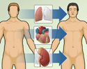
Heart transplant - overview
Animation
-
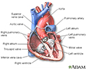
Heart, section through the middle - illustration
The interior of the heart is composed of valves, chambers, and associated vessels.
Heart, section through the middle
illustration
-
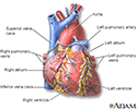
Heart, front view - illustration
The external structures of the heart include the ventricles, atria, arteries and veins. Arteries carry blood away from the heart while veins carry blood into the heart. The vessels colored blue indicate the transport of blood with relatively low content of oxygen and high content of carbon dioxide. The vessels colored red indicate the transport of blood with relatively high content of oxygen and low content of carbon dioxide.
Heart, front view
illustration
-
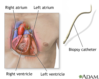
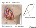
Biopsy catheter - illustration
When a small piece of heart muscle tissue is needed for examination, a heart biopsy can be performed. A catheter is carefully threaded into an artery or vein to gain access into the heart. A bioptome (catheter with jaws in its tip) is then introduced. Once the bioptome is in place, three to five small pieces of tissue from the heart muscle are removed. The test is performed routinely after heart transplantation to detect potential rejection. It may also be performed when cardiomyopathy, myocarditis, cardiac amyloidosis, or other disorders are suspected.
Biopsy catheter
illustration
-
Heart, section through the middle - illustration
The interior of the heart is composed of valves, chambers, and associated vessels.
Heart, section through the middle
illustration
-
Heart, front view - illustration
The external structures of the heart include the ventricles, atria, arteries and veins. Arteries carry blood away from the heart while veins carry blood into the heart. The vessels colored blue indicate the transport of blood with relatively low content of oxygen and high content of carbon dioxide. The vessels colored red indicate the transport of blood with relatively high content of oxygen and low content of carbon dioxide.
Heart, front view
illustration
-
Biopsy catheter - illustration
When a small piece of heart muscle tissue is needed for examination, a heart biopsy can be performed. A catheter is carefully threaded into an artery or vein to gain access into the heart. A bioptome (catheter with jaws in its tip) is then introduced. Once the bioptome is in place, three to five small pieces of tissue from the heart muscle are removed. The test is performed routinely after heart transplantation to detect potential rejection. It may also be performed when cardiomyopathy, myocarditis, cardiac amyloidosis, or other disorders are suspected.
Biopsy catheter
illustration
Review Date: 5/5/2016
Reviewed By: Michael A. Chen, MD, PhD, Associate Professor of Medicine, Division of Cardiology, Harborview Medical Center, University of Washington Medical School, Seattle, WA. Also reviewed by David Zieve, MD, MHA, Isla Ogilvie, PhD, and the A.D.A.M. Editorial team.