Renal arteriography
Renal angiogram; Angiography - kidney; Renal angiography; Renal artery stenosis - arteriography
Renal arteriography is a special x-ray of the blood vessels of the kidneys.
How the Test is Performed
This test is done in the hospital. You will lie on an x-ray table.
Health care providers often use an artery near the groin for the test. Occasionally provider may use an artery in the wrist. Your provider will:
- Clean and shave the area.
- Apply a numbing medicine to the area.
- Place a needle into the artery.
- Pass a thin wire through the needle into the artery.
- Take out the needle.
- Insert a long, narrow, flexible tube called a catheter in its place.
The radiologist directs the catheter into correct position using x-ray images of the body. An instrument called fluoroscope sends the images to a TV monitor, which the provider can see.
The catheter is pushed ahead over the wire into the aorta (main blood vessel from the heart). It then enters into the kidney artery. The test uses a special dye (called contrast) to help the arteries show up on the x-ray. The blood vessels of the kidneys are not seen with ordinary x-rays. The dye flows through the catheter into the kidney artery.
X-ray images are taken as the dye moves through the blood vessels. Saline (sterile salt water) containing a blood thinner may also be sent through the catheter to keep blood in the area from clotting.
The catheter is removed after the x-rays are taken. A closure device is placed in the groin or pressure is applied to the area to stop the bleeding. The area is checked after 10 or 15 minutes and a bandage is applied. You may be asked to keep your leg straight for 4 to 6 hours after the procedure.
How to Prepare for the Test
Tell the provider if:
- You are pregnant
- You have ever had any bleeding problems
- You currently take blood thinners, including daily aspirin
-
You ever had any
allergic reactions
, especially those related to x-ray contrast material or iodine substances
Allergic reactions
Allergic reactions are sensitivities to substances called allergens that come into contact with the skin, nose, eyes, respiratory tract, and gastroin...
 ImageRead Article Now Book Mark Article
 - You have ever been diagnosed with kidney failure or poorly functioning kidneys
You must sign a consent form. DO NOT eat or drink anything for 6 to 8 hours before the test. You will be given a hospital gown to wear and asked to remove all jewelry. You may be given a pain pill (sedative) before the procedure.
How the Test will Feel
You will lie flat on the x-ray table. There is usually a cushion but it is not as comfortable as a bed. You may feel a sting when the anesthesia medicine is given. You will not feel the dye. You may feel some pressure and discomfort as the catheter is positioned.
Some people feel a warm sensation when the dye is injected; however, most people cannot feel it. There may be slight tenderness and bruising at the site of the injection after the test.
Why the Test is Performed
Renal arteriography is often needed to help decide on the best treatment after other tests are done first. These include duplex ultrasound , CT abdomen, or a CT angiogram. These tests may show the following problems.
Duplex ultrasound
A duplex ultrasound is a test to see how blood moves through your arteries and veins.

-
Abnormal widening of an artery, called an
aneurysm
Aneurysm
An aneurysm is an abnormal widening or ballooning of a part of an artery due to weakness in the wall of the blood vessel.
 ImageRead Article Now Book Mark Article
 -
Abnormal connections between veins and arteries (
fistulas
)
Fistulas
A fistula is an abnormal connection between 2 body parts, such as an organ or blood vessel and another structure. Fistulas are usually the result of...
 ImageRead Article Now Book Mark Article
 -
Blood clot blocking an artery supplying the kidney
Blood clot blocking an artery supplying...
Acute arterial occlusion of the kidney is a sudden, severe blockage of the artery that supplies blood to the kidney.
 ImageRead Article Now Book Mark Article
 -
Unexplained high blood pressure thought to be due to
narrowing of the blood vessels of the kidneys
Narrowing of the blood vessels of the k...
Renovascular hypertension is high blood pressure due to narrowing of the arteries that carry blood to the kidneys. This condition is also called ren...
 ImageRead Article Now Book Mark Article
 - Benign tumors and cancers involving the kidneys
- Active bleeding from the kidney
This test is often used to examine donors and recipients before a kidney transplant. The result determines the number of arteries and veins on each kidney.
Normal Results
Results may vary. Talk to your doctor about the meaning of your specific test results.
What Abnormal Results Mean
Renal angiography may show the presence of tumors, narrowing of the artery or aneurysms (widening of the vein or artery), blood clots, fistulas, or bleeding in the kidney.
The test may also be done with the following conditions:
-
Blockage of an artery by a blood clot
Blockage of an artery by a blood clot
Acute arterial occlusion of the kidney is a sudden, severe blockage of the artery that supplies blood to the kidney.
ImageRead Article Now Book Mark Article - Renal artery stenosis
-
Renal cell cancer
Renal cell cancer
Renal cell carcinoma is a type of kidney cancer that starts in the lining of very small tubes (tubules) in the kidney.
ImageRead Article Now Book Mark Article - Angiomyolipomas (noncancerous tumors of the kidney)
Some of these problems can be treated with techniques done at the same time the arteriogram is performed.
- Angioplasty is a procedure to open a narrowed or blocked blood vessels that supply blood to your kidneys.
- A stent is a small, metal mesh tube that keeps the artery open. It may be placed to keep a narrowed artery open
Risks
The procedure is generally safe. There may be some risks, such as:
- Allergic reaction to the dye (contrast medium)
- Arterial occlusion from dissection
- Damage to the artery or artery wall, which can lead to blood clots
- Kidney damage from damage to the artery or from the dye
There is low radiation exposure . Pregnant women and children are more sensitive to the risks related to x-rays.
Radiation exposure
Radiation sickness is illness and symptoms resulting from excessive exposure to ionizing radiation. There are two basic types of radiation: ionizing ...

Considerations
The test should NOT be done if you are pregnant or have severe bleeding problems.
Magnetic resonance angiography (MRA) or CT angiography (CTA) can be done instead. MRA and CTA are noninvasive and can provide similar imaging of the kidney arteries, although they cannot be used for treatment.
Magnetic resonance angiography
Magnetic resonance angiography (MRA) is an MRI exam of the blood vessels. Unlike traditional angiography that involves placing a tube (catheter) int...
References
Duddalwar VA, Jadvar H, Palmer SL, Boswell WD. Diagnostic kidney imaging. In: Skorecki K, Chertow GM, Marsden PA, MW Taal, Yu ASL, eds. Brenner and Rector's The Kidney . 10th ed. Philadelphia, PA: Elsevier Saunders; 2016:chap 28.
Mclafferty RB. Arteriography. In: Cronenwett JL, Johnston KW, eds. Rutherford's Vascular Surgery . 8th ed. Philadelphia, PA: Elsevier Saunders; 2014:chap 19.
Patel MS, Lumsden AB, Davies MG. Renal artery atheroembolism. In: Stanley JC, Veith FJ, Wakefield TW, eds. Current therapy in Vascular and Endovascular Surgery . 5th ed. Philadelphia, PA: Elsevier Saunders; 2014:811-812.
-
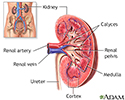
Kidney anatomy - illustration
The kidneys are responsible for removing wastes from the body, regulating electrolyte balance and blood pressure, and stimulating red blood cell production.
Kidney anatomy
illustration
-
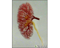
Kidney - blood and urine flow - illustration
This is the typical appearance of the blood vessels (vasculature) and urine flow pattern in the kidney. The blood vessels are shown in red and the urine flow pattern in yellow.
Kidney - blood and urine flow
illustration
-
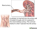
Renal arteries - illustration
A renal angiogram is a test used to examine the blood vessels of the kidneys. The test is performed by threading a catheter through the main vessel of the pelvis, up to the renal artery that leads into the kidney. Contrast medium is then injected into the renal artery through the catheter, and images of the vessels of the kidney are taken. The test is a useful aid in evaluating kidney function and diagnosing any narrowing of the arteries, blood clots, tumors or aneurysms.
Renal arteries
illustration
-
Kidney anatomy - illustration
The kidneys are responsible for removing wastes from the body, regulating electrolyte balance and blood pressure, and stimulating red blood cell production.
Kidney anatomy
illustration
-
Kidney - blood and urine flow - illustration
This is the typical appearance of the blood vessels (vasculature) and urine flow pattern in the kidney. The blood vessels are shown in red and the urine flow pattern in yellow.
Kidney - blood and urine flow
illustration
-
Renal arteries - illustration
A renal angiogram is a test used to examine the blood vessels of the kidneys. The test is performed by threading a catheter through the main vessel of the pelvis, up to the renal artery that leads into the kidney. Contrast medium is then injected into the renal artery through the catheter, and images of the vessels of the kidney are taken. The test is a useful aid in evaluating kidney function and diagnosing any narrowing of the arteries, blood clots, tumors or aneurysms.
Renal arteries
illustration
Review Date: 1/5/2016
Reviewed By: Jason Levy, MD, Northside Radiology Associates, Atlanta, GA. Also reviewed by David Zieve, MD, MHA, Isla Ogilvie, PhD, and the A.D.A.M. Editorial team.