Venogram - leg
Phlebogram - leg; Venography - leg; Angiogram - leg
Venography for legs is a test used to see the veins in the leg.
X-rays are a form of electromagnetic radiation, like visible light is. However, these rays are of higher energy. Therefore, they can go through the body to form an image on film. Structures that are dense (such as bone) will appear white, air will be black, and other structures will be shades of gray.
X-rays
X-rays are a type of electromagnetic radiation, just like visible light. An x-ray machine sends individual x-ray particles through the body. The im...

Veins are not normally seen in an x-ray, so a special dye is used to highlight them. This dye is called contrast.
How the Test is Performed
This test is usually done in a hospital. You will be asked to lie on an x-ray table. A numbing medicine is applied to the area. You may ask for a sedative if you are anxious about the test.
The health care provider places a needle into a vein in the foot of the leg being looked at. An intravenous (IV) line is inserted through the needle. The contrast dye flows through this line into the vein. A tourniquet may be placed on your leg so the dye flows into the deeper veins.
Intravenous
Intravenous means "within a vein. " Most often it refers to giving medicines or fluids through a needle or tube inserted into a vein. This allows th...
X-rays are taken as the dye flows through the leg.
The catheter is then removed, and the puncture site is bandaged.
How to Prepare for the Test
You will wear hospital clothing during this procedure. You will be asked to sign a consent form for the procedure. Remove all jewelry from the area being imaged.
Tell the provider:
- If you are pregnant
-
If you have
allergies to any medicines
Allergies to any medicines
Drug allergies are a group of symptoms caused by an allergic reaction to a drug (medicine).
 ImageRead Article Now Book Mark Article
 - Which medicines you are taking (including any herbal preparations)
-
If you have ever had any
allergic reactions
to x-ray contrast material or iodine substance
Allergic reactions
Allergic reactions are sensitivities to substances called allergens that come into contact with the skin, nose, eyes, respiratory tract, and gastroin...
 ImageRead Article Now Book Mark Article
How the Test will Feel
The x-ray table is hard and cold. You may want to ask for a blanket or pillow. You will feel a sharp poke when the intravenous catheter is inserted. As the dye is injected, you may experience a burning sensation.
There may be tenderness and bruising at the site of the injection after the test.
Why the Test is Performed
This test is used to identify and locate blood clots in the veins of the legs.
Blood clots
Blood clots are clumps that occur when blood hardens from a liquid to a solid. A blood clot that forms inside one of your veins or arteries is calle...

Normal Results
Free flow of the blood through the vein is normal.
What Abnormal Results Mean
Abnormal results may be due to a blockage. The blockage can be caused by:
-
Blood clot
Blood clot
Deep vein thrombosis (DVT) is a condition that occurs when a blood clot forms in a vein deep inside a part of the body. It mainly affects the large ...
 ImageRead Article Now Book Mark Article
 - Tumor
- Inflammation
Risks
Risks of this test are:
- Allergic reaction to the contrast dye
- Kidney failure, especially in the older adults or people with diabetes who take the medicine metformin (Glucophage)
- Worsening of a clot in the leg vein
There is low radiation exposure. However, most experts feel that the risk of most x-rays is smaller than other daily risks. Pregnant women and children are more sensitive to the risks of the x-ray.
Considerations
Ultrasound is used more often than this test because it has fewer risks and side effects. MRI and CT scans may also be used to look at the veins in the leg.
Ultrasound
Ultrasound uses high-frequency sound waves to make images of organs and structures inside the body.

References
Gillespie DL, Caliste XA. Venography. In: Cronenwett JL, Johnston KW, eds. Rutherford's Vascular Surgery . 8th ed. Philadelphia, PA: Elsevier Saunders; 2014:chap 20.
Ginsberg JS. Peripheral venous disease. In: Goldman L, Schafer AI, eds. Goldman's Cecil Medicine. 25th ed. Philadelphia, PA: Elsevier Saunders; 2016:chap 81.
-
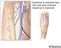
Leg venography - illustration
Leg venography is a procedure where contrast material is injected through a catheter in a vein to help visualize the internal structures by using x-rays. The test is used to identify and locate thrombi (blood clots) in the veins of the extremity that is affected.
Leg venography
illustration
-
Leg venography - illustration
Leg venography is a procedure where contrast material is injected through a catheter in a vein to help visualize the internal structures by using x-rays. The test is used to identify and locate thrombi (blood clots) in the veins of the extremity that is affected.
Leg venography
illustration
Review Date: 6/6/2016
Reviewed By: Deepak Sudheendra, MD, RPVI, Assistant Professor of Interventional Radiology & Surgery at the University of Pennsylvania Perelman School of Medicine, with an expertise in Vascular Interventional Radiology & Surgical Critical Care, Philadelphia, PA. Review provided by VeriMed Healthcare Network. Also reviewed by David Zieve, MD, MHA, Isla Ogilvie, PhD, and the A.D.A.M. Editorial team.