Histocompatibility antigen test
HLA typing; Tissue typing
A histocompatibility antigen blood test looks at proteins called human leukocyte antigens (HLAs). These are found on the surface of almost all cells in the human body. HLAs are found in large amounts on the surface of white blood cells. They help the immune system tell the difference between body tissue and substances that are not from your own body.
How the Test is Performed
Blood is drawn from a vein. In most cases a vein on the inside of the elbow or the back of the hand is used. First, the site where blood will be drawn is cleaned with germ-killing medicine (antiseptic). Then, the health care provider wraps an elastic band around the upper arm to apply pressure to the area and make the vein swell with blood.
Next, the provider gently inserts a needle into the vein. The blood collects into an airtight vial or tube attached to the needle. The elastic band is removed from your arm.
Once the blood has been collected, the needle is removed, and the puncture site is covered to stop any bleeding.
In infants or young children, a sharp tool called a lancet may be used to puncture the skin and make it bleed. The blood collects into a small glass tube called a pipette, or onto a slide or test strip. A bandage may be placed over the area if there is any bleeding.
How to Prepare for the Test
You do not need to prepare for this test.
How the Test Will Feel
You may feel slight pain or a sting when the needle is inserted. Afterward, there may be some throbbing.
Why the Test is Performed
The results from this test can be used to identify good matches for tissue grafts and organ transplants. These may include kidney transplant or bone marrow transplant .
Kidney transplant
A kidney transplant is surgery to place a healthy kidney into a person with kidney failure.

Bone marrow transplant
A bone marrow transplant is a procedure to replace damaged or destroyed bone marrow with healthy bone marrow stem cells. Bone marrow is the soft, fat...

It may also be used to:
- Diagnose certain autoimmune disorders
- Determine relationships between children and parents when such relationships are in question
- Monitor treatment with some medicines
Normal Results
You have a small set of HLAs that are passed down from your parents. Children, on average, will have half of their HLAs match half of their mother's and half of their HLAs match half of their father's.
It is unlikely that two unrelated people will have the same HLA makeup. However, identical twins may match each other.
Some HLA types are more common in certain autoimmune diseases . For example, HLA-B27 antigen is found in many people (but not all) with ankylosing spondylitis and Reiter syndrome .
Autoimmune diseases
An autoimmune disorder occurs when the body's immune system attacks and destroys healthy body tissue by mistake. There are more than 80 types of aut...

HLA-B27 antigen
HLA-B27 is a blood test to look for a protein that is found on the surface of white blood cells. The protein is called human leukocyte antigen B27 (...

Ankylosing spondylitis
Ankylosing spondylitis (AS) is a chronic form of arthritis. It mostly affects the bones and joints at the base of the spine where it connects with t...

Reiter syndrome
Reactive arthritis is a group of conditions that may involve the joints, eyes, and urinary and genital systems. These areas become swollen and infla...

Risks
Slight risks from having blood drawn may include:
- Excessive bleeding
- Fainting or feeling light-headed
- Hematoma (blood accumulating under the skin)
- Infection (a slight risk any time the skin is broken)
- Bruising
References
Wang, E. Human leukocyte antigen and human neutrophil antigen systems. In: Hoffman R, Benz EJ Jr, Silberstein LE, Heslop HE, Weitz JI, eds. Hematology: Basic Principles and Practice . 6th ed. Philadelphia, PA: Elsevier Saunders; 2012:chap 114.
-
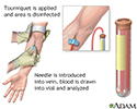
Blood test - illustration
Blood is drawn from a vein (venipuncture), usually from the inside of the elbow or the back of the hand. A needle is inserted into the vein, and the blood is collected in an air-tight vial or a syringe. Preparation may vary depending on the specific test.
Blood test
illustration
-
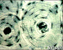
Bone Tissue - illustration
A photomicrograph of bone tissue. Bone tissue is obtained from a bone biopsy and examined under a microscope. This is a picture of how normal tissue appears when magnified.
Bone Tissue
illustration
-
Blood test - illustration
Blood is drawn from a vein (venipuncture), usually from the inside of the elbow or the back of the hand. A needle is inserted into the vein, and the blood is collected in an air-tight vial or a syringe. Preparation may vary depending on the specific test.
Blood test
illustration
-
Bone Tissue - illustration
A photomicrograph of bone tissue. Bone tissue is obtained from a bone biopsy and examined under a microscope. This is a picture of how normal tissue appears when magnified.
Bone Tissue
illustration
Review Date: 2/18/2015
Reviewed By: Frank A. Greco, MD, PhD, Director, Biophysical Laboratory, Edith Nourse Rogers Memorial Hospital, Bedford, MA. Review provided by VeriMed Healthcare Network. Also reviewed by David Zieve, MD, MHA, Isla Ogilvie, PhD, and the A.D.A.M. Editorial team.