Ultrasound
Sonogram
Ultrasound uses high-frequency sound waves to make images of organs and structures inside the body.
How the Test is Performed
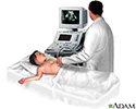
An ultrasound machine makes images so that organs inside the body can be examined. The machine sends out high-frequency sound waves, which reflect off body structures. A computer receives the waves and uses them to create a picture. Unlike with an x-ray or CT scan, this test does not use ionizing radiation.
The test is done in the ultrasound or radiology department.
- You will lie down for the test.
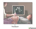
- A clear, water-based gel is applied to the skin on the area to be examined. The gel helps with the transmission of the sound waves.
- A handheld probe called a transducer is moved over the area being examined. You may need to change position so that other areas can be examined.
How to Prepare for the Test
Your preparation will depend on the part of the body being examined.
How the Test will Feel
Most of the time, ultrasound procedures do not cause discomfort. The conducting gel may feel a little cold and wet.
Why the Test is Performed
The reason for the test will depend on your symptoms. An ultrasound test may be used to identify problems involving:
-
Arteries in the neck
Arteries in the neck
Intravascular ultrasound (IVUS) is a diagnostic test. This test uses sound waves to see inside blood vessels. It is useful for evaluating the coron...
 ImageRead Article Now Book Mark Article
 -
Veins or arteries in the arms or legs
Veins or arteries in the arms or legs
This test uses ultrasound to look at the blood flow in the large arteries and veins in the arms and legs.
 ImageRead Article Now Book Mark Article
 -
Pregnancy
Pregnancy
A pregnancy ultrasound is an imaging test that uses sound waves to create a picture of how a baby is developing in the womb. It is also used to chec...
 ImageRead Article Now Book Mark Article
 -
Pelvis
Pelvis
A pelvic (transabdominal) ultrasound is an imaging test. It is used to examine organs in the pelvis.
Read Article Now Book Mark Article -
Abdomen and kidneys
Abdomen and kidneys
Abdominal ultrasound is a type of imaging test. It is used to look at organs in the abdomen, including the liver, gallbladder, spleen, pancreas, and...
 ImageRead Article Now Book Mark Article
 -
Breast
Breast
Breast ultrasound is a test that uses sound waves to examine the breasts.
 ImageRead Article Now Book Mark Article
 -
Thyroid
Thyroid
A thyroid ultrasound is an imaging method to see the thyroid, a gland in the neck that regulates metabolism.
 ImageRead Article Now Book Mark Article
 -
Eye and orbit
Eye and orbit
An eye and orbit ultrasound is a test to look at the eye area. It also measures the size and structures of the eye.
 ImageRead Article Now Book Mark Article
Normal Results
Results are considered normal if the organs and structures being examined look OK.
What Abnormal Results Mean
The meaning of abnormal results will depend on the part of the body being examined and the problem found. Talk to your health care provider about your questions and concerns.
Risks
There are no known risks. The test does not use ionizing radiation.
Considerations
Some types of ultrasound tests need to be done with a probe that is inserted into your body. Talk to your provider about how your test will be done.
References
Butts C. Ultrasound. In: Roberts JR, ed. Roberts and Hedges' Clinical Procedures in Emergency Medicine . 6th ed. Philadelphia, PA: Elsevier Saunders; 2014:chap 66.
Cosgrove DO, Eckersley RJ, Harvey CJ, Lim A. Ultrasound. In: Adam A, Dixon AK, Gillard JH, Schaefer-Prokop CM, eds. Grainger & Allison's Diagnostic Radiology: A Textbook of Medical Imaging . 6th ed. New York, NY: Elsevier; 2015:chap 3.
-
Abdominal ultrasound - illustration
Abdominal ultrasound is a scanning technique used to image the interior of the abdomen. Like the X-ray, MRI, and CT scan, it has its place as a diagnostic tool. Ultrasound scans use high frequency sound waves to produce an image and do not expose the individual to radiation. The procedure is painless and safe.
Abdominal ultrasound
illustration
-
Ultrasound in pregnancy - illustration
The ultrasound has become a standard procedure used during pregnancy. It can demonstrate fetal growth and can detect increasing numbers of conditions in the fetus including meningomyelocele, congenital heart disease, kidney abnormalities, hydrocephalus, anencephaly, club feet, and other deformities. Ultrasound does not produce ionizing radiation and is considered a very safe procedure for both the mother and the fetus.
Ultrasound in pregnancy
illustration
-
17 week ultrasound - illustration
During seventeen to twenty weeks of development, fetal movements known as quickening are commonly felt by the mother.
17 week ultrasound
illustration
-
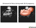
30 week ultrasound - illustration
Around 30 weeks, the growth of the brain markedly increases. Most systems are well developed, and the fetus can see and hear. A baby may survive if born this early, although the lungs may still be immature.
30 week ultrasound
illustration
-
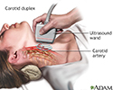
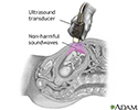
Carotid duplex - illustration
Carotid duplex is an ultrasound procedure performed to assess blood flow through the carotid artery to the brain. High-frequency sound waves are directed from a hand-held transducer probe to the area. These waves bounce off the arterial structures and produce a 2-dimensional image on a monitor, which will make obstructions or narrowing of the arteries visible.
Carotid duplex
illustration
-
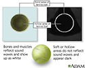
Ultrasound comparison - illustration
To demonstrate how an ultrasound works, imagine this tennis ball as an internal organ in the body. Like many organs, the tennis ball is solid on the outside and hollow on the inside. Solid structures, such as bones and muscles, reflect sound waves from the ultrasound transducer and show up as white in an ultrasound image. Soft or hollow areas, like chambers of the heart, do not reflect sound waves and appear as black. The white ring is the outer edge of the tennis ball being reflected back as an image while the center hollow area remains as black.
Ultrasound comparison
illustration
-
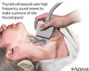
Thyroid ultrasound - illustration
Thyroid ultrasound is a sound wave picture of the thyroid gland taken by a hand-held instrument and translated to a 2-dimensional picture on a monitor. It is used in diagnosis of tumors, cysts or goiters of the thyroid, and is a painless, no-risk procedure.
Thyroid ultrasound
illustration
-
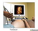
Ultrasound - illustration
Ultrasound is a scanning technique used to image the growing fetus. The transducer portion emits inaudible sound waves, which fan out as they travel through your abdomen. When they hit dense structures like the fetus and the wall of your uterus, the sound waves bounce back to the transducer and are translated into a visual image by the computer.
Ultrasound
illustration
-
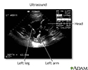
Ultrasound, normal fetus- ventricles of brain - illustration
This is a normal fetal ultrasound performed at 17 weeks gestation. The development of the brain and nervous system begins early in fetal development. During an ultrasound, the technician usually looks for the presence of brain ventricles. Ventricles are spaces in the brain that are filled with fluid. In this early ultrasound, the ventricles can be seen as light lines extending through the skull, seen in the upper right side of the image.
Ultrasound, normal fetus- ventricles of brain
illustration
-
3D ultrasound - illustration
3D ultrasound provides a three dimensional image of the fetus. Sound waves are sent at different angles by the transducer for the computer to reconstruct the height, width, and depth of the image.
3D ultrasound
illustration

-
Abdominal ultrasound - illustration
Abdominal ultrasound is a scanning technique used to image the interior of the abdomen. Like the X-ray, MRI, and CT scan, it has its place as a diagnostic tool. Ultrasound scans use high frequency sound waves to produce an image and do not expose the individual to radiation. The procedure is painless and safe.
Abdominal ultrasound
illustration
-
Ultrasound in pregnancy - illustration
The ultrasound has become a standard procedure used during pregnancy. It can demonstrate fetal growth and can detect increasing numbers of conditions in the fetus including meningomyelocele, congenital heart disease, kidney abnormalities, hydrocephalus, anencephaly, club feet, and other deformities. Ultrasound does not produce ionizing radiation and is considered a very safe procedure for both the mother and the fetus.
Ultrasound in pregnancy
illustration
-
17 week ultrasound - illustration
During seventeen to twenty weeks of development, fetal movements known as quickening are commonly felt by the mother.
17 week ultrasound
illustration
-
30 week ultrasound - illustration
Around 30 weeks, the growth of the brain markedly increases. Most systems are well developed, and the fetus can see and hear. A baby may survive if born this early, although the lungs may still be immature.
30 week ultrasound
illustration
-
Carotid duplex - illustration
Carotid duplex is an ultrasound procedure performed to assess blood flow through the carotid artery to the brain. High-frequency sound waves are directed from a hand-held transducer probe to the area. These waves bounce off the arterial structures and produce a 2-dimensional image on a monitor, which will make obstructions or narrowing of the arteries visible.
Carotid duplex
illustration
-
Ultrasound comparison - illustration
To demonstrate how an ultrasound works, imagine this tennis ball as an internal organ in the body. Like many organs, the tennis ball is solid on the outside and hollow on the inside. Solid structures, such as bones and muscles, reflect sound waves from the ultrasound transducer and show up as white in an ultrasound image. Soft or hollow areas, like chambers of the heart, do not reflect sound waves and appear as black. The white ring is the outer edge of the tennis ball being reflected back as an image while the center hollow area remains as black.
Ultrasound comparison
illustration
-
Thyroid ultrasound - illustration
Thyroid ultrasound is a sound wave picture of the thyroid gland taken by a hand-held instrument and translated to a 2-dimensional picture on a monitor. It is used in diagnosis of tumors, cysts or goiters of the thyroid, and is a painless, no-risk procedure.
Thyroid ultrasound
illustration
-
Ultrasound - illustration
Ultrasound is a scanning technique used to image the growing fetus. The transducer portion emits inaudible sound waves, which fan out as they travel through your abdomen. When they hit dense structures like the fetus and the wall of your uterus, the sound waves bounce back to the transducer and are translated into a visual image by the computer.
Ultrasound
illustration
-
Ultrasound, normal fetus- ventricles of brain - illustration
This is a normal fetal ultrasound performed at 17 weeks gestation. The development of the brain and nervous system begins early in fetal development. During an ultrasound, the technician usually looks for the presence of brain ventricles. Ventricles are spaces in the brain that are filled with fluid. In this early ultrasound, the ventricles can be seen as light lines extending through the skull, seen in the upper right side of the image.
Ultrasound, normal fetus- ventricles of brain
illustration
-
3D ultrasound - illustration
3D ultrasound provides a three dimensional image of the fetus. Sound waves are sent at different angles by the transducer for the computer to reconstruct the height, width, and depth of the image.
3D ultrasound
illustration
Review Date: 7/3/2016
Reviewed By: Jason Levy, MD, Northside Radiology Associates, Atlanta, GA. Also reviewed by David Zieve, MD, MHA, Isla Ogilvie, PhD, and the A.D.A.M. Editorial team.