Ventricular septal defect
VSD; Interventricular septal defect; Congenital heart defect - VSD
Ventricular septal defect is a hole in the wall that separates the right and left ventricles of the heart. Ventricular septal defect is one of the most common congenital (present from birth) heart defects. It may occur by itself or with other congenital diseases.
Causes
Before a baby is born, the right and left ventricles of its heart are not separate. As the fetus grows, a wall forms to separate these 2 ventricles. If the wall does not completely form, a hole remains. This hole is known as a ventricular septal defect, or a VSD.
Ventricular septal defect is a common congenital heart defect. The baby may have no symptoms and the hole can close over time as the wall continues to grow after birth. If the hole is large, too much blood will be pumped to the lungs. This can lead to heart failure.
The cause of VSD is not yet known. This defect often occurs along with other congenital heart defects.
In adults, ventricular septal defects are a rare but serious complication of heart attacks. These holes do not result from a birth defect.
Symptoms
People with ventricular septal defects may not have symptoms. However, if the hole is large, the baby often has symptoms related to heart failure.
The most common symptoms include:
- Shortness of breath
- Fast breathing
- Hard breathing
- Paleness
- Failure to gain weight
- Fast heart rate
- Sweating while feeding
- Frequent respiratory infections
Exams and Tests
Listening with a stethoscope most often reveals a heart murmur. The loudness of the murmur is related to the size of the defect and amount of blood crossing the defect.
Tests may include:
-
Cardiac catheterization
(rarely needed, unless there are concerns of high blood pressure in the lungs)
Cardiac catheterization
Cardiac catheterization involves passing a thin flexible tube (catheter) into the right or left side of the heart. The catheter is most often insert...
 ImageRead Article Now Book Mark Article
 -
Chest x-ray
: looks to see if there is a large heart with fluid in the lungs
Chest x-ray
A chest x-ray is an x-ray of the chest, lungs, heart, large arteries, ribs, and diaphragm.
 ImageRead Article Now Book Mark Article
 -
ECG
: shows signs of an enlarged left ventricle
ECG
An electrocardiogram (ECG) is a test that records the electrical activity of the heart.
 ImageRead Article Now Book Mark Article
 -
Echocardiogram
: used to make a definite diagnosis
Echocardiogram
An echocardiogram is a test that uses sound waves to create pictures of the heart. The picture and information it produces is more detailed than a s...
 ImageRead Article Now Book Mark Article
 - MRI of the heart: used to find out how much blood is getting to the lungs
Treatment
If the defect is small, no treatment may be needed. But the baby should be closely monitored by a health care provider. This is to make sure that the hole eventually closes properly and signs of heart failure do not occur.
Babies with a large VSD who have symptoms related to heart failure may need medicine to control the symptoms and surgery to close the hole. Medicines may include digitalis (digoxin) and diuretics.
If symptoms continue, even with medicine, surgery to close the defect with a patch is needed. Some VSDs can be closed with a special device during a cardiac catheterization , which avoids the need for surgery. However, only certain types of defects can successfully be treated this way.
Catheterization
Cardiac catheterization involves passing a thin flexible tube (catheter) into the right or left side of the heart. The catheter is most often insert...
Having surgery for a VSD with no symptoms is controversial. Discuss this carefully with your provider.
Outlook (Prognosis)
Many small defects will close on their own. Surgery can repair defects that do not close. In most cases, a person will not have any ongoing medical issues related to the defect if it is closed with surgery or closes on its own. Complications may occur if a large defect is not treated.
Possible Complications
Complications may include:
-
Aortic insufficiency
(leaking of the valve that separates the left ventricle from the aorta)
Aortic insufficiency
Aortic insufficiency is a heart valve disease in which the aortic valve does not close tightly. This allows blood to flow from the aorta (the larges...
 ImageRead Article Now Book Mark Article
 - Damage to the electrical conduction system of the heart during surgery (causing an irregular heart rhythm)
-
Delayed growth and development (
failure to thrive
in infancy)
Failure to thrive
Failure to thrive refers to children whose current weight or rate of weight gain is much lower than that of other children of similar age and gender....
Read Article Now Book Mark Article -
Heart failure
Heart failure
Heart failure is a condition in which the heart is no longer able to pump oxygen-rich blood to the rest of the body efficiently. This causes symptom...
 ImageRead Article Now Book Mark Article
 - Infective endocarditis (bacterial infection of the heart)
-
Pulmonary hypertension
(high blood pressure in the lungs) leading to failure of the right side of the heart
Pulmonary hypertension
Pulmonary hypertension is high blood pressure in the arteries of the lungs. It makes the right side of the heart work harder than normal.
 ImageRead Article Now Book Mark Article
When to Contact a Medical Professional
Most often, this condition is diagnosed during routine exam of an infant. Call your infant's provider if the baby seems to be having trouble breathing, or if the baby seems to have an unusual number of respiratory infections.
Prevention
Except for VSD that is caused by a heart attack, this condition is always present at birth.
Drinking alcohol and using the antiseizure medicines depakote and dilantin during pregnancy may increase the risk for VSDs. Other than avoiding these things during pregnancy, there is no known way to prevent a VSD.
References
Fraser CD, Carberry KE. Congenital heart disease. In: Townsend CM Jr, Beauchamp RD, Evers BM, Mattox KL, eds. Sabiston Textbook of Surgery . 19th ed. Philadelphia, PA: Elsevier Saunders; 2012:chap 59.
Webb GD, Smallhorn JF, Therrien J, Redington AN. Congenital heart disease. In: Mann DL, Zipes DP, Libby P, Bonow RO, Braunwald E, eds. Braunwald's Heart Disease: A Textbook of Cardiovascular Medicine . 10th ed. Philadelphia, PA: Elsevier Saunders; 2015:chap 62.
-
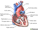
Heart, section through the middle - illustration
The interior of the heart is composed of valves, chambers, and associated vessels.
Heart, section through the middle
illustration
-
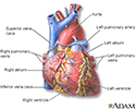
Heart, front view - illustration
The external structures of the heart include the ventricles, atria, arteries and veins. Arteries carry blood away from the heart while veins carry blood into the heart. The vessels colored blue indicate the transport of blood with relatively low content of oxygen and high content of carbon dioxide. The vessels colored red indicate the transport of blood with relatively high content of oxygen and low content of carbon dioxide.
Heart, front view
illustration
-
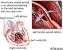
Ventricular septal defect - illustration
Ventricular septal defect is a congenital defect of the heart, that occurs as an abnormal opening in the wall that separates the right and left ventricles. Ventricular septal defect may also be associated with other heart defects. Many small defects will close on their own. For those defects that do not spontaneously close, the outcome is good with surgical repair.
Ventricular septal defect
illustration
-
Heart, section through the middle - illustration
The interior of the heart is composed of valves, chambers, and associated vessels.
Heart, section through the middle
illustration
-
Heart, front view - illustration
The external structures of the heart include the ventricles, atria, arteries and veins. Arteries carry blood away from the heart while veins carry blood into the heart. The vessels colored blue indicate the transport of blood with relatively low content of oxygen and high content of carbon dioxide. The vessels colored red indicate the transport of blood with relatively high content of oxygen and low content of carbon dioxide.
Heart, front view
illustration
-
Ventricular septal defect - illustration
Ventricular septal defect is a congenital defect of the heart, that occurs as an abnormal opening in the wall that separates the right and left ventricles. Ventricular septal defect may also be associated with other heart defects. Many small defects will close on their own. For those defects that do not spontaneously close, the outcome is good with surgical repair.
Ventricular septal defect
illustration
Review Date: 10/22/2015
Reviewed By: Larry A. Weinrauch, MD, Assistant Professor of Medicine, Harvard Medical School, Cardiovascular Disease and Clinical Outcomes Research, Watertown, MA. Review provided by VeriMed Healthcare Network. Also reviewed by David Zieve, MD, MHA, Isla Ogilvie, PhD, and the A.D.A.M. Editorial team.