Ectropion
Ectropion is the turning out of the eyelid so that the inner surface is exposed. It most often affects the lower eyelid.
Causes
Ectropion is very often caused by the aging process. The connective (supporting) tissue of the eyelid becomes weak. This causes the lid to turn out so that the side of the lower lid is no longer against the eyeball. It can also be caused by:
-
A defect that occurs before birth (for example, in children with
Down syndrome
)
Down syndrome
Down syndrome is a genetic condition in which a person has 47 chromosomes instead of the usual 46.
Read Article Now Book Mark Article -
Facial palsy
Facial palsy
Bell palsy is a disorder of the nerve that controls movement of the muscles in the face. This nerve is called the facial or seventh cranial nerve. D...
 ImageRead Article Now Book Mark Article
 - Scar tissue from burns
Symptoms
Symptoms include:
- Dry, painful eyes
-
Excess
tearing of the eye
(
epiphora
)
Tearing of the eye
Watery eyes means you have too many tears draining from the eyes. Tears help keep the surface of the eye moist. They wash away particles and foreig...
 ImageRead Article Now Book Mark Article
Epiphora
Watery eyes means you have too many tears draining from the eyes. Tears help keep the surface of the eye moist. They wash away particles and foreig...
ImageRead Article Now Book Mark Article - Eyelid turns outward (downward)
-
Long-term (chronic)
conjunctivitis
Conjunctivitis
The conjunctiva is a clear layer of tissue lining the eyelids and covering the white of the eye. Conjunctivitis occurs when the conjunctiva becomes...
 ImageRead Article Now Book Mark Article
 -
Keratitis
Keratitis
Interstitial keratitis is inflammation of the tissue of the cornea, the clear window on the front of the eye. The condition can lead to vision loss....
ImageRead Article Now Book Mark Article - Redness of the lid and white part of the eye
If you have ectropion, you will most likely have excess tearing. This happens because the eye gets dry, then makes more tears. The excess tears can't get into the tear drainage duct. Therefore, they build up inside the lower lid and then spill over the edge of the lid onto the cheek.
Exams and Tests
The health care provider will make a diagnosis by doing an exam of the eyes and eyelids. Special tests are not needed most of the time.
Treatment
Artificial tears (a lubricant) may ease dryness and keep the cornea moist. Ointment may be helpful when the eye can't close all of the way, such as when you are asleep. Surgery is very often effective. The surgeon will tighten the muscles that hold the eyelids in place. It may be done as outpatient surgery setting. A medicine is used to numb the area (local anesthesia) before the surgery.
Outlook (Prognosis)
The outcome very often good with treatment.
Possible Complications
Corneal dryness and irritation may lead to:
-
Corneal abrasions
Corneal abrasions
Corneal injury is a wound to the part of the eye known as the cornea. The cornea is the crystal clear (transparent) tissue that covers the front of ...
 ImageRead Article Now Book Mark Article
 -
Corneal ulcers
Corneal ulcers
The cornea is the clear tissue at the front of the eye. A corneal ulcer is an open sore in the outer layer of the cornea. It is often caused by inf...
ImageRead Article Now Book Mark Article - Eye infections
Corneal ulcers can cause vision loss.
When to Contact a Medical Professional
Call your provider if you have symptoms of ectropion.
If you have ectropion, get emergency medical help if you have:
- Vision that is getting worse
- Pain
-
Sensitivity to light
Sensitivity to light
Photophobia is eye discomfort in bright light.
ImageRead Article Now Book Mark Article - Eye redness that is getting worse quickly
Prevention
Most cases are cannot be prevented. You may use artificial tears or ointments to prevent injury to the cornea.
References
Belliveau MJ, Vargason CW, Burkat CN, Marcet MM, Belliveau MJ, Goel S. Ectropion. American Academy of Ophthalmology Web site. Updated November 17, 2015. eyewiki.aao.org/Ectropion#Etiology . Accessed September 8, 2016.
Cahill KV, Doxanas MT. Eyelid abnormalities: ectropion, entropion, trichiasis. In: Tasman W, Jaeger EA, eds. Duane's Ophthalmology 2013 ed . Philadelphia, PA: Lippincott Williams & Wilkins; 2013:vol 5, chap 73.
Robinson FO, Richard J, Collin O. Ectropion. In: Yanoff M, Duker JS, eds. Ophthalmology . 4th ed. Philadelphia, PA: Elsevier Saunders; 2014:chap 12.7.
Yanoff M, Cameron DL. Diseases of the visual system. In: Goldman L, Schafer AI, eds. Goldman-Cecil Medicine . 25th ed. Philadelphia, PA: Elsevier Saunders; 2016:chap 431.
-
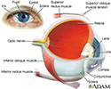
Eye - illustration
The eye is the organ of sight, a nearly spherical hollow globe filled with fluids (humors). The outer layer or tunic (sclera, or white, and cornea) is fibrous and protective. The middle tunic layer (choroid, ciliary body and the iris) is vascular. The innermost layer (the retina) is nervous or sensory. The fluids in the eye are divided by the lens into the vitreous humor (behind the lens) and the aqueous humor (in front of the lens). The lens itself is flexible and suspended by ligaments which allow it to change shape to focus light on the retina, which is composed of sensory neurons.
Eye
illustration
-
Eye - illustration
The eye is the organ of sight, a nearly spherical hollow globe filled with fluids (humors). The outer layer or tunic (sclera, or white, and cornea) is fibrous and protective. The middle tunic layer (choroid, ciliary body and the iris) is vascular. The innermost layer (the retina) is nervous or sensory. The fluids in the eye are divided by the lens into the vitreous humor (behind the lens) and the aqueous humor (in front of the lens). The lens itself is flexible and suspended by ligaments which allow it to change shape to focus light on the retina, which is composed of sensory neurons.
Eye
illustration
Review Date: 8/20/2016
Reviewed By: Franklin W. Lusby, MD, ophthalmologist, Lusby Vision Institute, La Jolla, CA. Also reviewed by David Zieve, MD, MHA, Isla Ogilvie, PhD, and the A.D.A.M. Editorial team.