Choroidal dystrophies
Choroideremia; Gyrate atrophy; Central areolar choroidal dystrophy
Choroidal dystrophy is an eye disorder that involves a layer of blood vessels called the choroid . These vessels are between the sclera and retina .
Choroid
The choroid is the layer of blood vessels and connective tissue between the white of the eye and retina (at the back of the eye). It is part of the ...

Sclera
The sclera is the white outer coating of the eye. It is tough, fibrous tissue that extends from the cornea (the clear front section of the eye) to t...
Retina
The retina is the light-sensitive layer of tissue at the back of the eyeball. Images that come through the eye's lens are focused on the retina. Th...
In most cases, choroidal dystrophy is due to an abnormal gene, which is passed down through families. It most often affects males, starting in childhood.
The first symptoms are peripheral vision loss and vision loss at night. An eye surgeon who specializes in the retina (back of the eye) can diagnose this disorder.
Exams and Tests
The following tests may be needed to diagnose the condition:
-
Electroretinography
Electroretinography
Electroretinography is a test to measure the electrical response of the eye's light-sensitive cells, called rods and cones. These cells are part of ...
 ImageRead Article Now Book Mark Article
 -
Fluorescein angiography
Fluorescein angiography
Fluorescein angiography is an eye test that uses a special dye and camera to look at blood flow in the retina and choroid. These are the two layers ...
 ImageRead Article Now Book Mark Article
 - Genetic testing
References
Genead MA, Fishman GA, Gover S. Hereditary choroidal diseases. In: Ryan SJ, Sadda SR, Hinton DR, Schachat AP, et al, eds. Retina . 5th ed. Philadelphia, PA: Elsevier Saunders; 2013:chap 43.
Grover S, Fishman GA, Ganead MA. Choroidal dystrophies In: Yanoff M, Duker JS, eds. Ophthalmology. 4th ed. Philadelphia, PA: Elsevier Saunders; 2014:chap 6.15.
-
External and internal eye anatomy - illustration
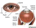
The cornea allows light to enter the eye. As light passes through the eye the iris changes shape by expanding and letting more light through or constricting and letting less light through to change pupil size. The lens then changes shape to allow the accurate focusing of light on the retina. Light excites photoreceptors that eventually, through a chemical process, transmit nerve signals through the optic nerve to the brain. The brain processes these nerve impulses into sight.
External and internal eye anatomy
illustration
-
External and internal eye anatomy - illustration
The cornea allows light to enter the eye. As light passes through the eye the iris changes shape by expanding and letting more light through or constricting and letting less light through to change pupil size. The lens then changes shape to allow the accurate focusing of light on the retina. Light excites photoreceptors that eventually, through a chemical process, transmit nerve signals through the optic nerve to the brain. The brain processes these nerve impulses into sight.
External and internal eye anatomy
illustration
Review Date: 11/4/2015
Reviewed By: Franklin W. Lusby, MD, ophthalmologist, Lusby Vision Institute, La Jolla, CA. Also reviewed by David Zieve, MD, MHA, Isla Ogilvie, PhD, and the A.D.A.M. Editorial team.