Coal worker's pneumoconiosis
Black lung disease; Pneumoconiosis; Anthrosilicosis
Coal worker's pneumoconiosis is a lung disease that results from breathing in dust from coal, graphite, or man-made carbon over a long time.
Causes
Coal worker's pneumoconiosis occurs in two forms: simple and complicated (also called progressive massive fibrosis, or PMF).
Your risk of developing coal worker's pneumoconiosis depends on how long you have been around coal dust. Most people with this disease are older than 50. Smoking does not increase your risk of developing this disease, but it may have an added harmful effect on the lungs.
If coal worker's pneumoconiosis occurs with rheumatoid arthritis , it is called Caplan syndrome .
Rheumatoid arthritis
Rheumatoid arthritis (RA) is a long-term disease. It leads to inflammation of the joints and surrounding tissues. It can also affect other organs....

Caplan syndrome
Caplan syndrome is swelling (inflammation) and scarring of the lungs. It occurs in people with rheumatoid arthritis who have breathed in mining dust...

Symptoms
Symptoms of this condition include:
-
Cough
Cough
Coughing is an important way to keep your throat and airways clear. But too much coughing may mean you have a disease or disorder. Some coughs are d...
 ImageRead Article Now Book Mark Article
 -
Shortness of breath
Shortness of breath
Breathing difficulty may involve:Difficult breathingUncomfortable breathingFeeling like you are not getting enough air
ImageRead Article Now Book Mark Article - Coughing up of black sputum
Exams and Tests
The doctor will do a physical exam and listen to your lungs with a stethoscope. A chest x-ray or chest CT scan will be performed. You will also likely need lung function tests.
Chest x-ray
A chest x-ray is an x-ray of the chest, lungs, heart, large arteries, ribs, and diaphragm.

Chest CT scan
A chest CT (computed tomography) scan is an imaging method that uses x-rays to create cross-sectional pictures of the chest and upper abdomen....

Treatment
Treatment may include any of the following, depending on how severe your symptoms are:
- Medicines to keep the airways open and reduce mucus
- Pulmonary rehabilitation to help you learn ways to breathe better
- Oxygen therapy
Support Groups
Ask your health care provider about Black Lung Clinics in your area. Information can be found at the National Coalition of Black Lung and Respiratory Disease Clinics website: blacklungcoalition.org/clinics .
Outlook (Prognosis)
Outcome for the simple form is usually good. It rarely causes disability or death. The complicated form may cause shortness of breath that worsens over time.
Possible Complications
Complications may include:
-
Chronic
bronchitis
Bronchitis
Acute bronchitis is swelling and inflamed tissue in the main passages that carry air to the lungs. This swelling narrows the airways, which makes it...
ImageRead Article Now Book Mark Article -
Chronic obstructive pulmonary disease
(COPD)
Chronic obstructive pulmonary disease
Chronic obstructive pulmonary disease (COPD) is a common lung disease. Having COPD makes it hard to breathe. There are two main forms of COPD:Chroni...
 ImageRead Article Now Book Mark Article
 -
Cor pulmonale
(failure of the right side of the heart)
Cor pulmonale
Cor pulmonale is a condition that causes the right side of the heart to fail. Long-term high blood pressure in the arteries of the lung and right ve...
 ImageRead Article Now Book Mark Article
 -
Respiratory failure
Respiratory failure
Respiratory acidosis is a condition that occurs when the lungs cannot remove all of the carbon dioxide the body produces. This causes body fluids, e...
ImageRead Article Now Book Mark Article
Prevention
Wear a protective mask when working around coal, graphite, or man-made carbon. Companies should enforce the maximum permitted dust levels. Avoid smoking.
References
Cowie RL, Becklake MR. Pneumoconioses. In: Broaddus VC, Mason RJ, Ernst JD, et al, eds. Murray and Nadel's Textbook of Respiratory Medicine . 6th ed. Philadelphia, PA: Elsevier Saunders; 2016:chap 73.
Tarlo SM. Occupational lung disease. In: Goldman L, Schafer AI, eds. Goldman's Cecil Medicine . 25th ed. Philadelphia, PA: Elsevier Saunders; 2016:chap 93.
-
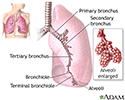
Lungs - illustration
The major features of the lungs include the bronchi, the bronchioles and the alveoli. The alveoli are the microscopic blood vessel-lined sacks in which oxygen and carbon dioxide gas are exchanged.
Lungs
illustration
-
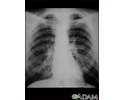
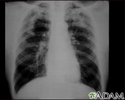
Coal worker's lungs - chest X-ray - illustration
This chest x-ray shows coal worker's lungs. There are diffuse, small, light areas on both sides (1 to 3 mm) in all parts of the lungs. Diseases that may result in an x-ray like this include: simple coal workers pneumoconiosis (CWP) - stage I, simple silicosis, miliary tuberculosis, histiocytosis X (eosinophilic granuloma), and other diffuse infiltrate pulmonary diseases.
Coal worker's lungs - chest X-ray
illustration
-
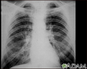
Coal workers pneumoconiosis - stage II - illustration
This chest x-ray shows stage II coal worker's pneumoconiosis (CWP). There are diffuse, small light areas on both sides of the lungs. Other diseases that may explain these x-ray findings include simple silicosis, disseminated tuberculosis, metastatic lung cancer, and other diffuse, infiltrative pulmonary diseases.
Coal workers pneumoconiosis - stage II
illustration
-
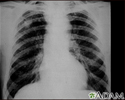
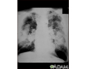
Coal workers pneumoconiosis - stage II #2 - illustration
This chest x-ray shows coal workers pneumoconiosis - stage II. There are diffuse, small (2 to 4 mm each), light areas throughout both lungs. In the right upper lung (seen on the left side of the picture), there is a light area (measuring approximately 2 cm by 4 cm) with poorly defined borders, representing coalescence (merging together) of previously distinct light areas. Diseases which may explain these x-ray findings include simple coal workers pneumoconiosis (CWP) - stage II, silico-tuberculosis, disseminated tuberculosis, metastatic lung cancer, and other diffuse infiltrative pulmonary diseases.
Coal workers pneumoconiosis - stage II #2
illustration
-
Coal workers pneumoconiosis, complicated - illustration
This picture shows complicated coal workers pneumoconiosis. There are diffuse, small, light areas (3 to 5 mm) in all areas on both sides of the lungs. There are large light areas which run together with poorly defined borders in the upper areas on both sides of the lungs. Diseases which may explain these X-ray findings include complicated coal workers pneumoconiosis (CWP), silico-tuberculosis, disseminated tuberculosis, metastatic lung cancer, and other diffuse infiltrative pulmonary diseases.
Coal workers pneumoconiosis, complicated
illustration
-
Coal workers pneumoconiosis, complicated #2 - illustration
This picture shows complicated coal workers pneumoconiosis. There are diffuse, massive light areas that run together in the upper and middle parts of both lungs. These are superimposed on a background of small and poorly distinguishable light areas that are diffuse and located in both lungs. Diseases which may explain these x-ray findings include, but are not limited to: complicated coal workers pneumoconiosis (CWP), silico-tuberculosis, and metastatic lung cancer.
Coal workers pneumoconiosis, complicated #2
illustration
-
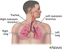
Respiratory system - illustration
Air is breathed in through the nasal passageways, travels through the trachea and bronchi to the lungs.
Respiratory system
illustration
-
Lungs - illustration
The major features of the lungs include the bronchi, the bronchioles and the alveoli. The alveoli are the microscopic blood vessel-lined sacks in which oxygen and carbon dioxide gas are exchanged.
Lungs
illustration
-
Coal worker's lungs - chest X-ray - illustration
This chest x-ray shows coal worker's lungs. There are diffuse, small, light areas on both sides (1 to 3 mm) in all parts of the lungs. Diseases that may result in an x-ray like this include: simple coal workers pneumoconiosis (CWP) - stage I, simple silicosis, miliary tuberculosis, histiocytosis X (eosinophilic granuloma), and other diffuse infiltrate pulmonary diseases.
Coal worker's lungs - chest X-ray
illustration
-
Coal workers pneumoconiosis - stage II - illustration
This chest x-ray shows stage II coal worker's pneumoconiosis (CWP). There are diffuse, small light areas on both sides of the lungs. Other diseases that may explain these x-ray findings include simple silicosis, disseminated tuberculosis, metastatic lung cancer, and other diffuse, infiltrative pulmonary diseases.
Coal workers pneumoconiosis - stage II
illustration
-
Coal workers pneumoconiosis - stage II #2 - illustration
This chest x-ray shows coal workers pneumoconiosis - stage II. There are diffuse, small (2 to 4 mm each), light areas throughout both lungs. In the right upper lung (seen on the left side of the picture), there is a light area (measuring approximately 2 cm by 4 cm) with poorly defined borders, representing coalescence (merging together) of previously distinct light areas. Diseases which may explain these x-ray findings include simple coal workers pneumoconiosis (CWP) - stage II, silico-tuberculosis, disseminated tuberculosis, metastatic lung cancer, and other diffuse infiltrative pulmonary diseases.
Coal workers pneumoconiosis - stage II #2
illustration
-
Coal workers pneumoconiosis, complicated - illustration
This picture shows complicated coal workers pneumoconiosis. There are diffuse, small, light areas (3 to 5 mm) in all areas on both sides of the lungs. There are large light areas which run together with poorly defined borders in the upper areas on both sides of the lungs. Diseases which may explain these X-ray findings include complicated coal workers pneumoconiosis (CWP), silico-tuberculosis, disseminated tuberculosis, metastatic lung cancer, and other diffuse infiltrative pulmonary diseases.
Coal workers pneumoconiosis, complicated
illustration
-
Coal workers pneumoconiosis, complicated #2 - illustration
This picture shows complicated coal workers pneumoconiosis. There are diffuse, massive light areas that run together in the upper and middle parts of both lungs. These are superimposed on a background of small and poorly distinguishable light areas that are diffuse and located in both lungs. Diseases which may explain these x-ray findings include, but are not limited to: complicated coal workers pneumoconiosis (CWP), silico-tuberculosis, and metastatic lung cancer.
Coal workers pneumoconiosis, complicated #2
illustration
-
Respiratory system - illustration
Air is breathed in through the nasal passageways, travels through the trachea and bronchi to the lungs.
Respiratory system
illustration
Review Date: 6/22/2015
Reviewed By: Denis Hadjiliadis, MD, MHS, Associate Professor of Medicine, Pulmonary, Allergy, and Critical Care, Perelman School of Medicine, University of Pennsylvania, Philadelphia, PA. Also reviewed by David Zieve, MD, MHA, Isla Ogilvie, PhD, and the A.D.A.M. Editorial team.